. 2011 Sep;301(3):F520-8.
doi: 10.1152/ajprenal.00075.2011. Epub 2011 May 25.

Interaction of H+ with the extracellular and intracellular aspects of hMATE1

Affiliations

Interaction of H+ with the extracellular and intracellular aspects of hMATE1

Yodying Dangprapai et al. Am J Physiol Renal Physiol. 2011 Sep.

Abstract

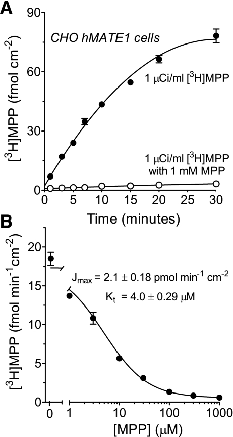
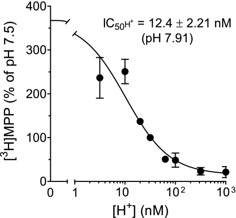
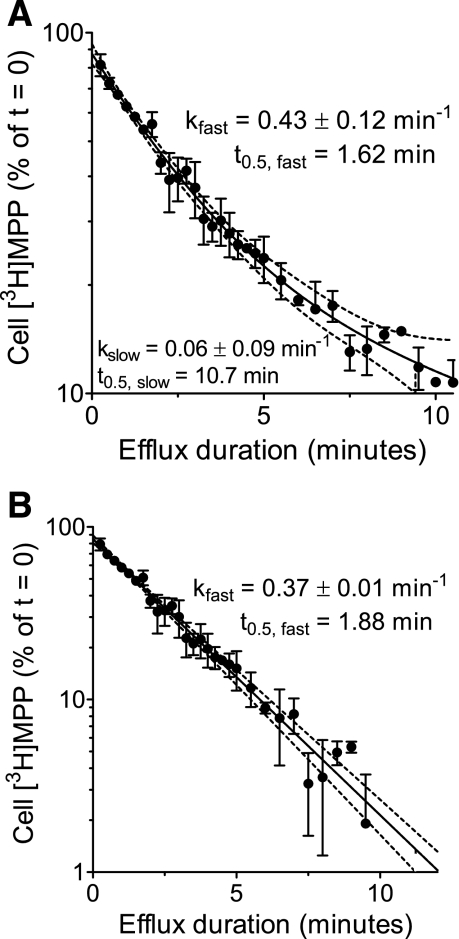
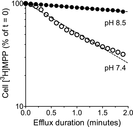
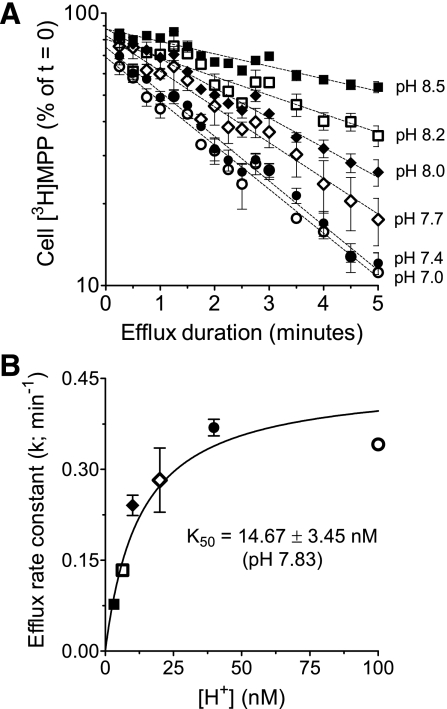
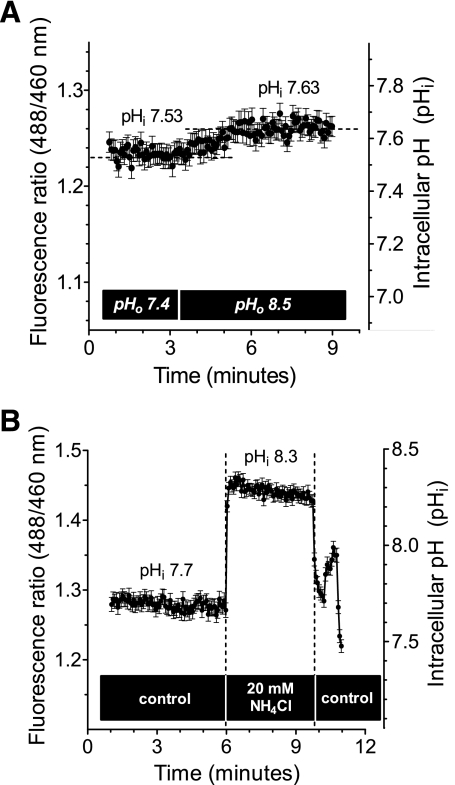
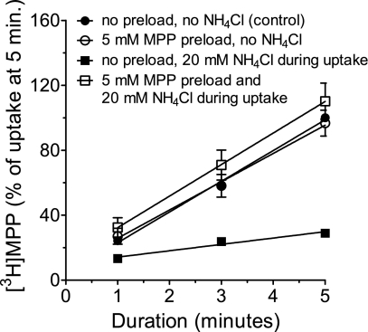
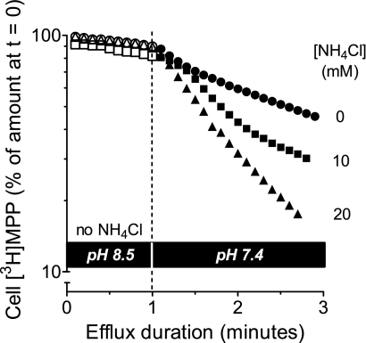
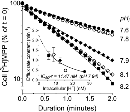
Human multidrug and toxin extrusion 1 (hMATE1, SLC47A1) is a major candidate for being the molecular identity of organic cation/proton (OC/H(+)) exchange activity in the luminal membrane of renal proximal tubules. Although physiological function of hMATE1 supports luminal OC efflux, the kinetics of hMATE1-mediated OC transport have typically been characterized through measurement of uptake, i.e., the interaction between outward-facing hMATE1 and OCs. To examine kinetics of hMATE1-mediated transport in a more physiologically relevant direction, i.e., an interaction between inward-facing hMATE1 and cytoplasmic substrates, we measured the time course of hMATE1-mediated efflux of the prototypic MATE1 substrate, [(3)H]1-methyl-4-phenylpyridinium, under a variety of intra- and extracellular pH conditions, from Chinese hamster ovary cells that stably expressed the transporter. In this study, we showed that an IC(50)/K(i) for interaction between extracellular H(+) and outward-facing hMATE1 determined from conventional uptake experiments [12.9 ± 1.23 nM (pH 7.89); n = 9] and from the efflux protocol [14.7 ± 3.45 nM (pH 7.83); n = 3] was not significantly different (P = 0.6). Furthermore, kinetics of interaction between intracellular H(+) and inward-facing hMATE1 determined using the efflux protocol revealed an IC(50) for H(+) of 11.5 nM (pH 7.91), consistent with symmetrical interactions of H(+) with the inward-facing and outward-facing aspects of hMATE1.


Figures

Fig. 1.
Fig. 1.
Time course and kinetics of [3H]MPP uptake in Chinese hamster ovary (CHO) human multidrug and toxin extrusion 1 (hMATE1) cells. A: [3H]MPP accumulated linearly in CHO hMATE1 cells up to ∼10 min (·). [3H]MPP uptake was inhibited by coincubating the cells with 1 mM nonlabeled MPP (○). Buffer pH was 8.5. Each point is the mean (±SE) of uptake measured in 2 wells of a 24-well plate. B: uptake (5 min; pH 8.5) of [3H]MPP transport measured in the presence of increasing concentrations of unlabeled MPP (from which the kinetics of MPP transport were determined). Data shown are from a single representative experiment. Each point is the mean (±SE) of uptakes measured in 3 wells of a 24-well plate.
Fig. 2.
Fig. 2.
IC50 of extracellular H+ as an inhibitor of hMATE1-mediated MPP uptake. Five-minute uptakes of 13 nM [3H]MPP into CHO hMATE1 cells were plotted as a function of extracellular [H+]. Each point is the mean (±SE) of uptakes determined in 5 separate experiments (n = 5).
Fig. 3.
Fig. 3.
Time course of MPP loss from CHO hMATE1 cells. A: following a 20-min preload of 13.5 nM [3H]MPP, the amount of labeled MPP remaining in the cells was measured as a function of time after efflux was initiated in an MPP-free buffer of pH 7.4 ([H+]o = 39.8 nM). Each point is the mean [3H]MPP content (±SE) of cells in 2 wells of a 48-well plate in a single representative experiment. The semilog plot reveals that there were multiple compartments involved in [3H]MPP efflux from CHO hMATE1 cells as discussed in the text. The solid line represents 2-phase exponential decline in cell [3H]MPP (kinetic parameters listed in the figure). B: time course of the first-order (single phase) decline in cell [3H]MPP, reflecting correction for sequestered [3H]MPP (see text for discussion). The dotted lines in both graphs represent the 95% confidence interval of the fitted values (solid line).
Fig. 4.
Fig. 4.
Time course of [2-(4-nitro-2,1,3-benzoxadiazol-7-yl)aminoethyl]trimethylammonium (NBD-TMA) efflux in CHO hMATE1 cells. CHO hMATE1 cells were preloaded with 0.5 mM NBD-TMA at 37°C for 15 min. Following aspiration of the medium, the cells were transferred to a fluorescent microscope, rinsed with Waymouth buffer (WB) at pH 8.5 at which point an image was collected representing t = 0. Subsequent images were collected at the indicated time points following introduction of test media. For each condition, the cells were illuminated only during collection of the image. The 10-min images were from different fields of cells (to avoid the influence of photobleaching).
Fig. 5.
Fig. 5.
Effect of extracellular H+ on hMATE1-mediated [3H]MPP efflux. Efflux was performed at extracellular pH 8.5 ([H+]out = 3.2 nM) and at pH 7.4 ([H+]out = 39.8 nM) using the efflux chamber protocol.
Fig. 6.
Fig. 6.
Effect of increasing extracellular [H+] on the kinetics of hMATE1-mediated [3H]MPP efflux. A: time course of [3H]MPP efflux from the “fast” cytoplasmic compartment (as defined in the text) was determined in buffers of decreasing external pH. The value of the slow compartment used for each condition was determined from the efflux profile at pHo 7.0. Each point is the mean of results obtained in 3 separate experiments. B: rate constants for [3H]MPP efflux from the fast compartment (the slopes of the lines fit to the data in A) plotted as a function of extracellular [H+] (line fit using Michaelis-Menten equation).
Fig. 7.
Fig. 7.
Intracellular pH (pHi) in CHO hMATE1 cells. A: representative experiment showing the modest change in pHi associated with acute change of pHo; in this case, a shift from pHo of 7.4 to pHo of 8.5 increased pHi from 7.53 to 7.63. B: upon acute exposure to 20 mM NH4Cl, the pHi of CHO hMATE1 cells increased rapidly (within 8 s) from a baseline pHi value (pHi of 7.57, i.e., [H+]i = 27.1 ± 1.79 nM; n = 21) to a new value (pHi of 8.2, i.e., [H+]i = 5.8 ± 0.35 nM; n = 4) that was relatively stable for at least 3 min. Data shown are from a single representative experiment. (Each point of the fluorescence ratio in both A and B was an average value measured from 28 CHO hMATE1 cells.)
Fig. 8.
Fig. 8.
Effect of NH4Cl and MPP preload on hMATE1-mediated [3H]MPP uptake. The time course of hMATE1-mediated uptake of [3H]MPP (13.5 nM) was measured under the conditions indicated in the figure. In every case, pHo was 8.5. Each point is the mean (±SE) of uptake values (measured in triplicate) from 3 separate experiments (n = 3).
Fig. 9.
Fig. 9.
Effect of NH4Cl on hMATE1-mediated [3H]MPP efflux. Cells were preloaded with [3H]MPP (36.8 nM) for 15 min. Efflux into a buffer of pH 8.5 was measured for 1 min, at which point the cells were exposed to a buffer of pH 7.4 containing the indicated concentrations of NH4Cl (to decrease [H+]i). Efflux into these new solutions was then determined for 2 min. Single representative examples are shown for each condition.
Fig. 10.
Fig. 10.
Kinetics of interaction between inward-facing hMATE1 and intracellular H+. The time course of hMATE1-mediated [3H]MPP efflux was measured for each of the indicated values of intracellular pH. Each point is the mean of values determined in 3–6 separate experiments. Inset: relationship between cytoplasmic [H+] and the calculated rate constants (means ± SE) for hMATE1-mediated [3H]MPP efflux.

References

    1. Anderson CMH, Thwaites DT. Hijacking solute carriers for proton-coupled drug transport. Physiology (Bethesda) 25: 364–377, 2010 - PubMed
    1. Bednarczyk D, Mash EA, Aavula BR, Wright SH. NBD-TMA: a novel fluorescent substrate of the peritubular organic cation transporter of renal proximal tubules. Pflügers Arch 440: 184–192, 2000 - PubMed
    1. Gorboulev V, Ulzheimer JC, Akhoundova A, Ulzheimer-Teuber I, Karbach U, Quester S, Baumann C, Lang F, Busch AE, Koepsell H. Cloning and characterization of two human polyspecific organic cation transporters. DNA Cell Biol 16: 871–881, 1997 - PubMed
    1. Groves CE, Evans KK, Dantzler WH, Wright SH. Peritubular organic cation transport in isolated rabbit proximal tubules. Am J Physiol Renal Fluid Electrolyte Physiol 266: F450–F458, 1994 - PubMed
    1. Guan L, Kaback HR. Lessons from lactose permease. Annu Rev Biophys Biomol Struct 35: 67–91, 2006 - PMC - PubMed

Publication types

LinkOut - more resources