

Total and regional brain volumes in a population-based normative sample from 4 to 18 years: the NIH MRI Study of Normal Brain Development
- PMID: 21613470
- PMCID: PMC3236790
- DOI: 10.1093/cercor/bhr018
Total and regional brain volumes in a population-based normative sample from 4 to 18 years: the NIH MRI Study of Normal Brain Development
Abstract
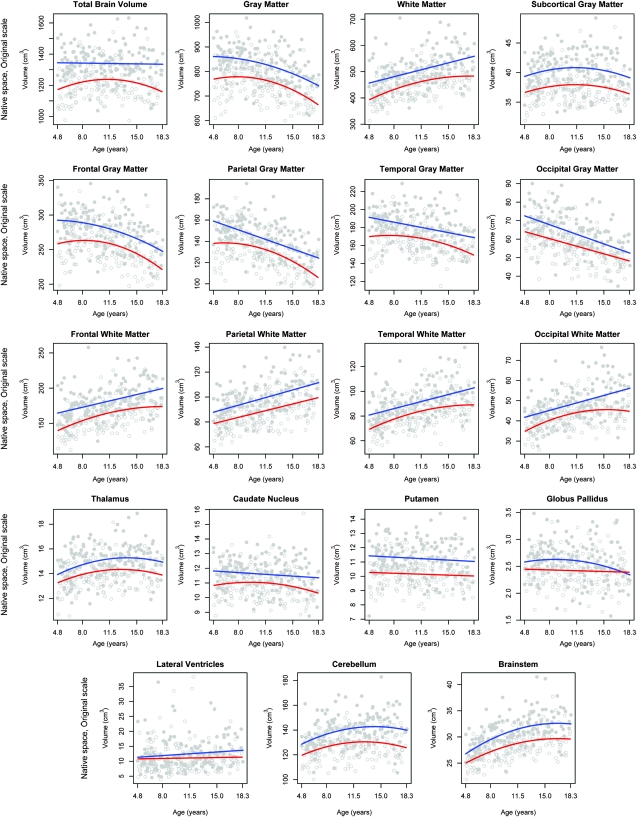
Using a population-based sampling strategy, the National Institutes of Health (NIH) Magnetic Resonance Imaging Study of Normal Brain Development compiled a longitudinal normative reference database of neuroimaging and correlated clinical/behavioral data from a demographically representative sample of healthy children and adolescents aged newborn through early adulthood. The present paper reports brain volume data for 325 children, ages 4.5-18 years, from the first cross-sectional time point. Measures included volumes of whole-brain gray matter (GM) and white matter (WM), left and right lateral ventricles, frontal, temporal, parietal and occipital lobe GM and WM, subcortical GM (thalamus, caudate, putamen, and globus pallidus), cerebellum, and brainstem. Associations with cross-sectional age, sex, family income, parental education, and body mass index (BMI) were evaluated. Key observations are: 1) age-related decreases in lobar GM most prominent in parietal and occipital cortex; 2) age-related increases in lobar WM, greatest in occipital, followed by the temporal lobe; 3) age-related trajectories predominantly curvilinear in females, but linear in males; and 4) small systematic associations of brain tissue volumes with BMI but not with IQ, family income, or parental education. These findings constitute a normative reference on regional brain volumes in children and adolescents.
Figures
References
-
- Akaike H. A new look at the statistical model identification. IEEE Trans Autom Contr. 1974;19:716–723.
-
- Almli CR, Rivkin MJ, McKinstry RC, Brain Development Cooperative Group The NIH MRI study of normal brain development (Objective-2): newborns, infants, toddlers, and preschoolers. Neuroimage. 2007;35:308–325. - PubMed
-
- Barrick TR, Mackay CE, Prima S, Maes F, Vandermeulen D, Crow TJ, Roberts N. Automatic analysis of cerebral asymmetry: an exploratory study of the relationship between brain torque and planum temporale asymmetry. Neuroimage. 2005;24:678–691. - PubMed
-
- Brain Development Cooperative Group (corresponding author Evans AC) The NIH MRI study of normal brain development. Neuroimage. 2006;30:184–202. - PubMed
-
- Caviness VS, Jr., Kennedy DN, Richelme C, Rademacher J, Filipek PA. The human brain age 7–11 years: a volumetric analysis based on magnetic resonance images. Cereb Cortex. 1996;6:726–736. - PubMed
Publication types
MeSH terms
Grants and funding
- N01-HD02-3343/HD/NICHD NIH HHS/United States
- N01-NS-9-2319/NS/NINDS NIH HHS/United States
- N01-NS-9-2315/NS/NINDS NIH HHS/United States
- NS34783/NS/NINDS NIH HHS/United States
- R01 NS034783/NS/NINDS NIH HHS/United States
- N01-NS-9-2317/NS/NINDS NIH HHS/United States
- N01-NS-9-2316/NS/NINDS NIH HHS/United States
- N01-NS-9-2320/NS/NINDS NIH HHS/United States
- N01-MH9-0002/MH/NIMH NIH HHS/United States
- N01-NS-9-2314/NS/NINDS NIH HHS/United States
- N01 HD023343/HD/NICHD NIH HHS/United States
- R56 NS034783/NS/NINDS NIH HHS/United States
LinkOut - more resources
Full Text Sources
Other Literature Sources
Medical
