. 2011 Jul 15;22(14):2436-47.
doi: 10.1091/mbc.E11-01-0001. Epub 2011 May 25.

Bundle-forming pilus retraction enhances enteropathogenic Escherichia coli infectivity

Affiliations

Bundle-forming pilus retraction enhances enteropathogenic Escherichia coli infectivity

Eitan E Zahavi et al. Mol Biol Cell. .

Abstract

Enteropathogenic Escherichia coli (EPEC) is an important human pathogen that causes acute infantile diarrhea. The type IV bundle-forming pili (BFP) of typical EPEC strains are dynamic fibrillar organelles that can extend out and retract into the bacterium. The bfpF gene encodes for BfpF, a protein that promotes pili retraction. The BFP are involved in bacterial autoaggregation and in mediating the initial adherence of the bacterium with its host cell. Importantly, BFP retraction is implicated in virulence in experimental human infection. How pili retraction contributes to EPEC pathogenesis at the cellular level remains largely obscure, however. In this study, an effort has been made to address this question using engineered EPEC strains with induced BFP retraction capacity. We show that the retraction is important for tight-junction disruption and, to a lesser extent, actin-rich pedestal formation by promoting efficient translocation of bacterial protein effectors into the host cells. A model is proposed whereby BFP retraction permits closer apposition between the bacterial and the host cell surfaces, thus enabling timely and effective introduction of bacterial effectors into the host cell via the type III secretion apparatus. Our studies hence suggest novel insights into the involvement of pili retraction in EPEC pathogenesis.


Figures
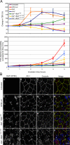
FIGURE 1:
FIGURE 1:
Induced BfpF expression perturbs the functions and morphology of TJs. MDCK cells cultured on semipermeable supports were infected with the indicated EPEC strains, or left untreated, as described in Materials and Methods. Infection with EPEC-bfpF +BfpF was performed in the absence (EPEC-bfpF +BfpFara−) or presence (EPEC-bfpF +BfpFara+) of arabinose. In some experiments, arabinose was added to the activation medium 15 min before cell infection and throughout the entire infection time (EPEC-bfpF +BfpFara+(T0)). In other experiments, cells were initially infected in the absence of arabinose for 2 h, and then arabinose was added to the infection medium for the rest of the infection time (EPEC-bfpF +BfpFara+(T2)). (A) Barrier functions. Changes in the TJ barrier functions were measured by tracking the alterations in TER (top panels) and monolayer permeability to FITC-dextran (bottom panels). Results are mean ± SE of at least three independent experiments, each of which was performed in duplicate. Data are normalized to values measured at time zero. Two-tailed Student's t test reveals that infection with EPEC expressing intact BfpF resulted in statistically significant reduction in TER and increase in cell monolayer permeability after 5 h of infection (see Table S4). (B) Junctional morphology. Polarized MDCK monolayers were infected with the indicated EPEC strains for 3 h at 37ºC, or left untreated. Cells were then fixed and costained with DAPI (bacteria and nuclei), anti–ZO-1 and anti-occludin antibodies. Fluorescence labeling was analyzed by confocal microscopy. Images are projections of x–y sections, taken along 2–3 μm from the cell's apex, where bacteria and junctional labeling was best visualized. Arrowheads indicate examples of breaks in occludin and ZO-1 staining. Bar = 10 μm.
FIGURE 2:
FIGURE 2:
BfpF expression is required for efficient translocationof EspF. HeLa cells were infected with the indicated EPEC strains or the pBAD24 vector as control (see Materials and Methods). All bacteria expressed the espF-blaM reporter gene. (A) Accumulation of cleaved CCF2 [P] (top panel) and its rate of accumulation [P′] (bottom panel) following EPEC infection. HeLa cells were infected with EPEC, and measurement of CCF2 product accumulation was carried out throughout the indicated times. Total product [P] and the rate of its accumulation [P′] were calculated, as explained in Materials and Methods. The dashed line in the bottom panel separates the two suggested phases of [P′] as a function of time (see Results and Table 1). The results are mean ± SE of four to eight measurements. (B) Levels of EPEC adherence to HeLa cells. HeLa cells were infected with EPEC for 30 or 60 min, and the number of adhered bacteria was determined as described in Materials and Methods and Figure S1. Data presented are mean ± SE of three independent experiments.
FIGURE 3:
FIGURE 3:
Induced BfpF expression is required for efficient production of actin-rich pedestals. (A) Visualization of bacteria and F-actin in fixed cells. MDCK cells were infected with the indicated EPEC strains for 45 and 120 min at 37ºC. Cells were fixed and stained with DAPI (bacteria and nuclei) and Texas-Red phalloidin (F- actin). Confocal images were acquired from the apical region of the cells and processed as indicated in Materials and Methods. Arrowheads point toward prominent F-actin accumulations associated with EPEC infection sites. Arrows and arrowheads with asterisks indicate weak or barely detectable F-actin accumulations at infection sites, respectively. Bar = 10 μm. (B and C) Quantitative time-lapse imaging analysis of GFP-actin (top panel) and mRFP-PM (bottom panel) accumulation at EPEC infection sites. Cells were cotransfected with plasmids encoding for GFP-actin and mRFP-PM. Cells were subsequently infected under the microscope, and time-lapse live imaging was performed (see Supplemental Movies 6–10). In the case where arabinose-mediated BfpF expression was tested, the inducer was added to the activation medium before cell infection [(EPEC-bfpF +BfpFara+(T0); (B)]; in another experiment, EPEC microcolonies were allowed to interact with the host cell surface for 15 min in medium lacking arabinose, and then the inducer was added until the end of the measurement [(EPEC-bfpF +BfpFara+(T15′); (C)]. Fluorescence intensity confined to EPEC attachment sites was quantitatively analyzed. The landing of EPEC microcolonies on the cell surface was identified by differential interference contrast microscopy, and data acquisition started at that time point (designated as time 0). Results presented in panel B are mean ± SE of four experiments. The experiment described in panel C (see Supplemental Movie 10) was repeated, but the microcolony was allowed to interact with the cell surface for 2 min in medium lacking arabinose (Supplemental Figure S3). (D) Ultrastructure of EPEC infection sites. Filter-cultured MDCK cells were infected with the indicated EPEC strains for 60 or 120 min at 37ºC. Cells were fixed and processed for observation by transmission electron microscopy, as described in Materials and Methods.
FIGURE 4:
FIGURE 4:
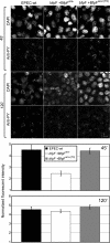
BfpF expression is required for efficient tyrosine phosphorylation of Tir in MDCK cells. MDCK cells were infected with EPEC for 45 or 120 min at 37°C. Cells were fixed and stained with DAPI to visualize bacteria and with anti-PY antibodies. Fluorescence was imaged by confocal microscopy, and representative images are shown in the top panel. The intensity of PY fluorescence levels associated with EPEC microcolonies was quantified, and data are presented in the bottom panel. Results are the mean ± SE of at least 20 infection sites imaged in two independent experiments.
FIGURE 5:
FIGURE 5:
BfpF expression is essential for efficient translocation and tyrosine phosphorylation of Tir in HeLa cells. (A) Tir translocation. HeLa cells were infected with EPEC-bfpF, EPEC-bfpF +BfpFara−, and EPEC-bfpF +BfpFara+(T0) expressing the tir-blaM reporter gene. Experiments were carried out and analyzed as indicated in Materials and Methods and Figure 2A. The results are mean ± SE of four to eight measurements. (B) Tir phosphorylation. HeLa cells were infected with the indicated EPEC strains or left untreated. Cells were lysed and subjected to immunoprecipitation (IP) with anti-PY antibodies followed by SDS–PAGE and immunoblotting (IB) with the same antibodies. PY-Tir is indicated with an arrow (top panel). Cell lysates were probed with anti–α-tubulin antibodies (middle panel, indicated with an arrow). Densitometric analysis was performed, and the PY-Tir/Tubulin ratio is shown (bottom panel). The results are representative of three independent experiments.
FIGURE 6:
FIGURE 6:
A working model linking pili retraction with the effectiveness of bacterial effector translocation. The model is described in the Discussion section.

References

    1. Allen-Vercoe E, Waddell B, Livingstone S, Deans J, DeVinney R. Enteropathogenic Escherichia coli Tir translocation and pedestal formation requires membrane cholesterol in the absence of bundle-forming pili. Cell Microbiol. 2006;8:613–624. - PubMed
    1. Anantha RP, Stone KD, Donnenberg MS. Role of BfpF, a member of the PilT family of putative nucleotide-binding proteins, in type IV pilus biogenesis and in interactions between enteropathogenic Escherichia coli and host cells. Infect Immun. 1998;66:122–131. - PMC - PubMed
    1. Biais N, Higashi DL, Brujic J, So M, Sheetz MP. Force-dependent polymorphism in type IV pili reveals hidden epitopes. Proc Natl Acad Sci USA. 2010;107:11358–11363. - PMC - PubMed
    1. Bieber D, Ramer SW, Wu CY, Murray WJ, Tobe T, Fernandez R, Schoolnik GK. Type IV pili, transient bacterial aggregates, and virulence of enteropathogenic Escherichia coli. Science. 1998;280:2114–2118. - PubMed
    1. Boettcher JP, Kirchner M, Churin Y, Kaushansky A, Pompaiah M, Thorn H, Brinkmann V, Macbeath G, Meyer TF. Tyrosine-phosphorylated caveolin-1 blocks bacterial uptake by inducing Vav2-RhoA-mediated cytoskeletal rearrangements. PLoS Biol. 2010;8(8):e1000457. - PMC - PubMed

Publication types