. 2011 Jul 15;22(14):2601-11.
doi: 10.1091/mbc.E11-02-0104. Epub 2011 May 25.

HOPS drives vacuole fusion by binding the vacuolar SNARE complex and the Vam7 PX domain via two distinct sites

Affiliations

HOPS drives vacuole fusion by binding the vacuolar SNARE complex and the Vam7 PX domain via two distinct sites

Lukas Krämer et al. Mol Biol Cell. .

Abstract

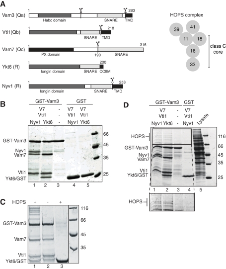
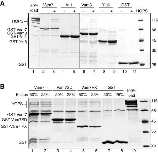
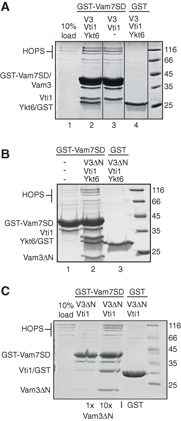
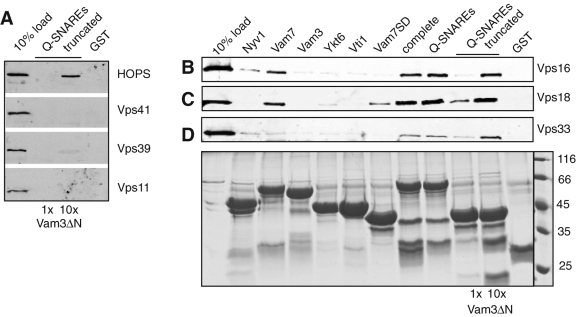
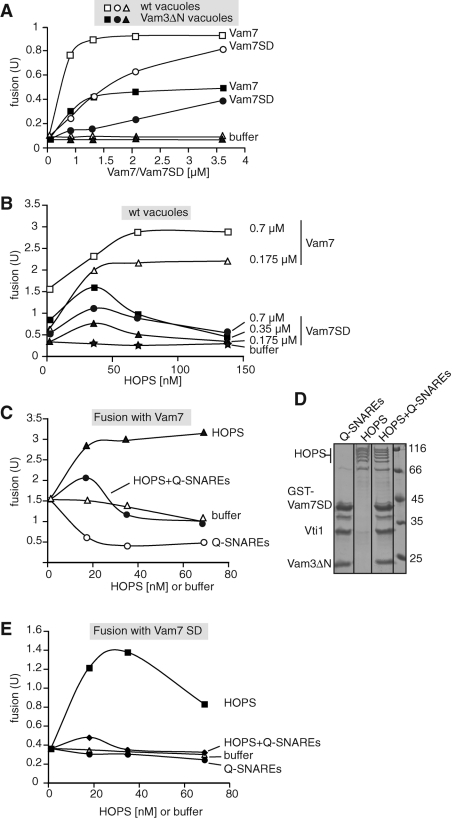
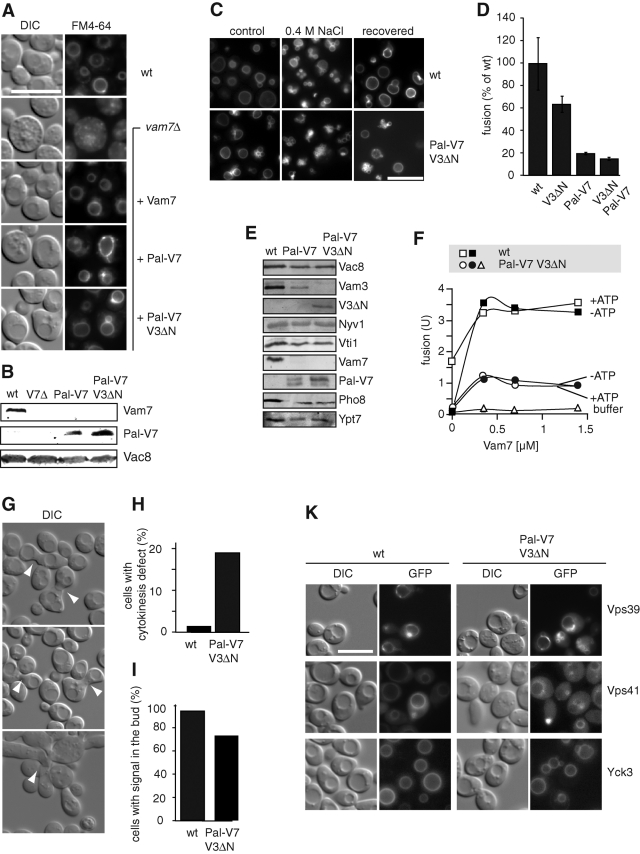
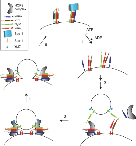
Membrane fusion within the endomembrane system follows a defined order of events: membrane tethering, mediated by Rabs and tethers, assembly of soluble N-ethylmaleimide-sensitive factor (NSF) attachment protein receptor (SNARE) complexes, and lipid bilayer mixing. Here we present evidence that the vacuolar HOPS tethering complex controls fusion through specific interactions with the vacuolar SNARE complex (consisting of Vam3, Vam7, Vti1, and Nyv1) and the N-terminal domains of Vam7 and Vam3. We show that homotypic fusion and protein sorting (HOPS) binds Vam7 via its subunits Vps16 and Vps18. In addition, we observed that Vps16, Vps18, and the Sec1/Munc18 protein Vps33, which is also part of the HOPS complex, bind to the Q-SNARE complex. In agreement with this observation, HOPS-stimulated fusion was inhibited if HOPS was preincubated with the minimal Q-SNARE complex. Importantly, artificial targeting of Vam7 without its PX domain to membranes rescued vacuole morphology in vivo, but resulted in a cytokinesis defect if the N-terminal domain of Vam3 was also removed. Our data thus support a model of HOPS-controlled membrane fusion by recognizing different elements of the SNARE complex.


Figures

FIGURE 1:
FIGURE 1:
Interaction of HOPS and the SNARE complex. (A, left) Schematic view of the domain structures of Vam3, Vti1, Vam7, Ykt6, and Nyv1. Scissors indicate the truncations used in this study. Membrane-anchoring domains/motifs are shown in black, the SNARE domain in gray. (A, right) Model of the HOPS complex. Subunits are arranged according to our previous findings (Ostrowicz et al., 2010). (B) Assembly of the SNARE complex. GST-tagged Vam3 or GST (10 μg each) were centrifuged for 30 min at 100,000 × g to remove aggregates and then preincubated with 12 μg of His-tagged SNAREs, which were pretreated similarly, for 2 h at 4°C, washed with buffer, and eluted in SDS-sample buffer. Eluted proteins were resolved on SDS–PAGE gels and stained with Coomassie. (C and D) Interaction of HOPS with assembled SNAREs. HOPS was added either as a purified complex (C) or from lysate of a strain overexpressing all HOPS subunits (D) to the SNARE complex or to GST. In (D), SNARE complexes with either Ykt6 or Nyv1 as the respective R-SNARE were used. Samples were incubated for 30 min with HOPS, then washed and eluted by boiling in SDS sample buffer. Proteins were analyzed as in (B).
FIGURE 2:
FIGURE 2:
Defining the interface between individual SNAREs and the HOPS complex. (A) Interaction of the individual SNAREs with HOPS. GST-tagged SNAREs and GST (12 μg) were incubated with or without purified HOPS complex and analyzed as before. (B) Interaction of Vam7 PX and the HOPS complex. Vam7, its SNARE domain (Vam7 SD), the PX domain (Vam7 PX), or GST was incubated with HOPS, and samples were processed as in Figure 1C. To estimate binding efficiency, 25 and 50% of the eluate were applied to the gel. It should be noted that binding of HOPS was most efficient to GST-Vam7 and GST-Vam7PX despite their lower protein amount on the beads.
FIGURE 3:
FIGURE 3:
Identification of a minimal SNARE complex that binds HOPS. (A) Interaction of HOPS with the Q-SNARE complex. GST-Vam7 SD or GST was incubated with Vam3, Vti1, and, where indicated, with Ykt6. The assembled complexes were then incubated for 30 min with purified HOPS and were washed. The bound proteins were eluted by boiling in sample buffer, resolved by SDS–PAGE, and stained with Coomassie. (B) Binding of HOPS to a minimal SNARE complex. Assembly was as in (A), except that a truncated Vam3 lacking the Habc domain (V3ΔN) was used. (C) Interaction of HOPS with a minimal Q-SNARE complex. Vam3ΔN was added in two concentrations to the SNAREs Vti1, GST-Vam7SD, and HOPS. After an incubation of 30 min with GSH beads, the beads were washed and eluted by boiling. The reactions were analyzed as in (A).
FIGURE 4:
FIGURE 4:
Two distinct binding sites within the HOPS for vacuolar SNAREs. GST-tagged SNAREs were either assembled in a minimal complex (GST-Vam7 SD, Vti1, and either one- or 10-fold excess of Vam3ΔN), or provided as individual proteins. HOPS or its subunits were purified as overproduced and TAP-tagged proteins from yeast, using IgG sepharose and TEV cleavage (see Materials and Methods and Ostrowicz et al., 2010). The purified proteins were then added to the beads, incubated for 30 min at 4°C, washed four times, and eluted by boiling in SDS sample buffer. A fraction (10%) of the eluate was then analyzed by SDS–PAGE and Western blotting against the calmodulin peptide present in the remaining tag of the HOPS subunits. In (A), HOPS, Vps41, Vps39, and Vps11 are shown. In (B), purified Vps16, (C) Vps18, or (D) Vps33 were added. The GST-tagged proteins or complexes (80%) were visualized by SDS–PAGE and Coomassie staining, and are shown (bottom) for Vps33. Control SDS–PAGE gels for all other pull-down assays were similar (unpublished data).
FIGURE 5:
FIGURE 5:
The two distinct HOPS binding sites can be distinguished in the vacuole fusion assay. (A) Analysis of wild-type and Vam3ΔN vacuoles in the fusion reaction. BJ and DKY vacuoles of the respective strains were purified and subjected to fusion as outlined in Materials and Methods. Reactions were carried out without ATP, but with increasing concentrations of Vam7 or Vam7 SNARE domain (Vam7 SD). Fusion reactions were developed after 90 min at 26°C. All fusion assays were carried out at least three times with similar results. (B) Cooperation of SNAREs and HOPS during fusion. Fusion of wild-type vacuoles was carried out in the presence of the indicated concentrations of Vam7 (0.7 and 0.176 μM) and Vam7 SNARE domain (SD; at 0.7, 0.35, and 0.175 μM) in the absence of ATP. HOPS was added in three different concentrations (35, 70, and 140 nM) to each Vam7-triggered fusion assay. (C–E) Competition of Q-SNAREs with HOPS. Q-SNAREs (GST-Vam7 SD, Vam3ΔN, and Vti1) were assembled on GSH beads, washed, and eluted with 15 mM reduced glutathione. Glutathione was removed from the complex in a SpinTrap G-25 column. One aliquot of the eluted SNAREs was incubated with HOPS for 2 h. A second aliquot was incubated with buffer as a control (D). HOPS, Q-SNAREs, or HOPS preincubated with Q-SNAREs (HOPS+Q-SNAREs) were then added at the indicated concentrations to wild-type vacuoles in the presence of Vam7 (0.7 μM) and absence of ATP (C). In (E), Vam7 SNARE domain (Vam7 SD) was added instead. All reactions were incubated for 90 min at 26°C and then developed.
FIGURE 6:
FIGURE 6:
Cooperation of the N-terminal domains of Vam3 and Vam7. (A) Vacuole morphology in cells lacking the Vam7 PX domain. Wild-type (wt) or vam7Δ cells with or without the indicated Vam7 variants were grown in YPD, stained with FM4–64, and visualized by fluorescence microscopy. In the bottom sample, Vam3 was truncated additionally in its N-terminal Habc domain. Scale bar = 10 μm. (B) Expression levels of Vam7 and Pal-Vam7. Proteins of wt, vam7Δ, Pal-Vam7ΔPX, and Pal-Vam7ΔPX Vam3ΔN cells were precipitated by trichloroacetic acid and separated by SDS–PAGE. Proteins were detected by Western blotting using specific antibodies. (C) In vivo fusion of cells with N-terminally truncated SNAREs. Wild-type cells and cells carrying Pal-Vam7ΔPX and Vam3ΔN were grown to logarithmic phase at 30°C, labeled with FM4–64, and then transferred to YPD with 0.4 M NaCl. After 10 min in salt, cells were reisolated and incubated in YPD for 1 h. Pictures were taken before salt stress (control), after 10 min in salt (0.4 M NaCl), and after 1 h in YPD (recovered). From 200 cells counted, the recovery rate of Pal-Vam7ΔPX Vam3ΔN cells was 95% of wild type. (D) Fusion of vacuoles with truncated SNAREs. Statistics of the fusion activity of wild-type vacuoles in comparison to mutants containing Vam3ΔN (V3ΔN), Pal-Vam7ΔPX (Pal-V7), or both are shown (n = 3). Optimal fusion conditions for all strains were used (0.7 μM Vam7 and 35 nM HOPS). (E) Expression level of selected vacuolar proteins. Purified vacuoles from the indicated strains were resolved on SDS–PAGE and blotted onto nitrocellulose membranes. Proteins were detected by Western blotting with the indicated antibodies. (F) Titration of Vam7 into the fusion reaction. Wild-type vacuoles and vacuoles with truncated Vam3 and Vam7 (V3ΔN Pal-V7) were incubated for 90 min at 26°C in the presence of 17.5 nM HOPS and increasing concentrations of full-length Vam7. ATP was added where indicated. Fusion reactions were then developed and analyzed as described in Materials and Methods. (G and H) Cells with N-terminally truncated Vam3 and Vam7 exhibit a cytokinesis defect. Cells were grown at 30°C in YPD and analyzed by microscopy using differential interference contrast (DIC) optics (G). Cells with defects in cytokinesis are indicated with arrowheads. Approximately 200 cells were counted for the statistical analysis (H). (I) Vacuole inheritance. The indicated cells were stained with 30 μM FM4–64 for 30 min, isolated, resuspended in fresh medium, and incubated for an additional 3 h. Vacuole staining in the daughter cell was scored as positive vacuole inheritance (n > 200 cells). (K) Localization of vacuole fusion proteins in mutant cells. The indicated proteins were N-terminally tagged with GFP and analyzed with fluorescence microscopy.
FIGURE 7:
FIGURE 7:
Model of the interplay of HOPS and SNAREs in the fusion cycle. Fusion is subdivided into five steps for simplicity: (1) Disassembly and activation of the assembled cis-SNAREs occurs in an ATP-dependent reaction. Under these conditions, Vam7 is released from the membrane. (2) Tethering of the vesicle with the target membrane. HOPS recruits Vam7 from the cytosol to the membrane and supports SNARE assembly. HOPS also protects the preassembled Q-SNAREs from disassembly by Sec18/Sec17. (3) Tethering of the vesicles or late endosome via the interaction of HOPS with Ypt7 and assembled SNAREs. (4) Fusion of the vesicle with the target membrane, which is mediated by trans-SNARE complexes. (5) Release of HOPS. SNAREs remain assembled and inactive as a cis-SNARE complex in the same membrane.

References

    1. Boeddinghaus C, Merz AJ, Laage R, Ungermann C. A cycle of Vam7p release from and PtdIns 3-P-dependent rebinding to the yeast vacuole is required for homotypic vacuole fusion. J Cell Biol. 2002;157:79–90. - PMC - PubMed
    1. Bowers K, Stevens TH. Protein transport from the late Golgi to the vacuole in the yeast Saccharomyces cerevisiae. Biochim Biophys Acta. 2005;1744:438–454. - PubMed
    1. Bracher A, Weissenhorn W. Structural basis for the Golgi membrane recruitment of Sly1p by Sed5p. EMBO J. 2002;21:6114–6124. - PMC - PubMed
    1. Brachmann CB, Davies A, Cost GJ, Caputo E, Li J, Hieter P, Boeke JD. Designer deletion strains derived from Saccharomyces cerevisiae S288C: a useful set of strains and plasmids for PCR-mediated gene disruption and other applications. Yeast. 1998;14:115–132. - PubMed
    1. Brett CL, Plemel RL, Lobinger BT, Vignali M, Fields S, Merz AJ. Efficient termination of vacuolar Rab GTPase signaling requires coordinated action by a GAP and a protein kinase. J Cell Biol. 2008;182:1141–1151. - PMC - PubMed

Publication types

MeSH terms

LinkOut - more resources