. 2011 Jun 14;108(24):9963-8.
doi: 10.1073/pnas.1012388108. Epub 2011 May 25.

Alternative mechanism for bacteriophage adsorption to the motile bacterium Caulobacter crescentus

Affiliations

Alternative mechanism for bacteriophage adsorption to the motile bacterium Caulobacter crescentus

Ricardo C Guerrero-Ferreira et al. Proc Natl Acad Sci U S A. .

Erratum in

  • Proc Natl Acad Sci U S A. 2011 Jul 19;108(29):12185

Abstract

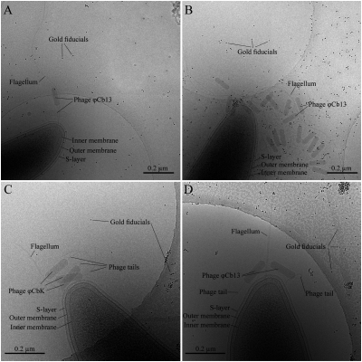
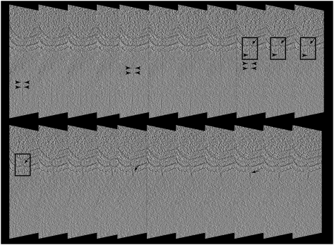
2D and 3D cryo-electron microscopy, together with adsorption kinetics assays of Cb13 and CbK phage-infected Caulobacter crescentus, provides insight into the mechanisms of infection. Cb13 and CbK actively interact with the flagellum and subsequently attach to receptors on the cell pole. We present evidence that the first interaction of the phage with the bacterial flagellum takes place through a filament on the phage head. This contact with the flagellum facilitates concentration of phage particles around the receptor (i.e., the pilus portals) on the bacterial cell surface, thereby increasing the likelihood of infection. Phage head filaments have not been well characterized and their function is described here. Phage head filaments may systematically underlie the initial interactions of phages with their hosts in other systems and possibly represent a widespread mechanism of efficient phage propagation.


Conflict of interest statement

The authors declare no conflict of interest.

Figures

Fig. 1.
Fig. 1.
Cryo-ET image, 3D reconstruction, and image analysis of ϕCb13 phages. (A) An image at 0° tilt of C. crescentus pole with two ϕCb13 phages attached. Fiducial gold particles are 10 nm in diameter. (B) A tomographic slice from Amira of the same region of the sample after tomographic reconstruction. (C) Same image after contrast inversion using the toolbox for 3D electron microscopy (EM) in Amira. (D) After thresholding with the island label module of the 3D EM toolbox the different regions are visualized. (E) Labels identify specific regions independently (i.e., each phage and the S-layer). Here, regions are visualized with different colors, yellow corresponding to the phages and green to the cell's S-layer. Arrowheads in E point to phage head filaments. The mouse cursor is used to select the labels to be included in the segmentation. Object selection is done through several slices and several times until all regions of interest are selected.
Fig. 2.
Fig. 2.
Automatically generated segmentations of ϕCb13-infected NA1000 C. crescentus cells. Notice the head filaments that extend from the phage and wrap around the flagellum. Orange-labeled densities inside yellow empty phages correspond to dislodged tail-head connectors. Full phages are depicted in blue and bacterial cell components in green. In A and B, the segmentation is superimposed over a Voltex (direct volume rendering where each point in the volume is assumed to emit and absorb light). Automatic segmentations were generated by using the IslandLabel module from the 3D-EM toolbox developed by Pruggnaller et al. (29).
Fig. 3.
Fig. 3.
Cryo-electron micrographs of plunge-frozen C. crescentus strains infected with either phage ϕCb13 or ϕCbK. (A) A NA1000 cell infected with phage ϕCb13. (B) A ΔmotA mutant cell infected with ϕCb13. Notice that flagellum-associated phage particles are oriented with their heads toward the flagellum. Phages attached to the cell pole are also observed. C and D correspond to ΔpilA mutants depicting successful adsorption of phage particles ϕCbK (C) and ϕCb13 (D) to the bacterial flagellum. Phage tails in C and D are not attached to the cell pole despite their close proximity. Scale bars 0.2 μm.
Fig. 4.
Fig. 4.
Image collage of the interaction between C. crescentus pilus and the tail of ϕCb13. Arrowheads point to the pilus filament in all images. (A) Slice through a tomogram of an infected NA1000 cell. The black rectangle highlights the area of pilus-phage interaction. (B) Segmented volume imposed in 3D over the same slice in A. Phages are depicted in blue, bacteria surface layer in green and pilus in orange. (C) Rotation of B to position the site of pilus-phage interaction in the front of the view. (D) Enlarged segmented volume.
Fig. 5.
Fig. 5.
Averaged 13-nm tomography slices of NA1000 C. crescentus cell infected with ϕCb13. Images are restricted to the cell pole and highlighted areas (rectangles) are shown where contact between the phage tail and cell pilus is evident. Arrowheads indicate pilus and arrows indicate portions of the phage tail.

References

    1. Schmidt JM, Stanier RY. Isolation and characterization of bacteriophages active against stalked bacteria. J Gen Microbiol. 1965;39:95–107. - PubMed
    1. Schade SZ, Adler J, Ris H. How bacteriophage chi attacks motile bacteria. J Virol. 1967;1:599–609. - PMC - PubMed
    1. Raimondo LM, Lundh NP, Martinez RJ. Primary adsorption site of phage PBS1: The flagellum of Bacillus subtilis. J Virol. 1968;2:256–264. - PMC - PubMed
    1. Lovett PS. PBPI: A flagella specific bacteriophage mediating transduction in Bacillus pumilus. Virology. 1972;47:743–752. - PubMed
    1. Edgar R, et al. Bacteriophage infection is targeted to cellular poles. Mol Microbiol. 2008;68:1107–1116. - PMC - PubMed

Publication types

MeSH terms

LinkOut - more resources