. 2011 Apr-Jun;7(2):140-4.
doi: 10.4161/org.7.2.16457. Epub 2011 Apr 1.

A rapid in vivo assay system for analyzing the organogenetic capacity of human kidney cells

Affiliations

A rapid in vivo assay system for analyzing the organogenetic capacity of human kidney cells

Tsahi Noiman et al. Organogenesis. 2011 Apr-Jun.

Abstract

Transplantation of human kidney-derived cells is a potential therapeutic modality for promoting regeneration of diseased renal tissue. However, assays that determine the ability of candidate populations for renal cell therapy to undergo appropriate differentiation and morphogenesis are limited. We report here a rapid and humane assay for characterizing tubulogenic potency utilizing the well-established chorioallantoic membrane CAM) of the chick embryo. Adult human kidney-derived cells expanded in monolayer were suspended in Matrigel and grafted onto the CAM. After a week, grafts were assessed histologically. Strikingly, many of the renal cells self-organized into tubular structures. Host blood vessels penetrated and presumably fed the grafts. Immuno- and histochemical staining revealed that tubular structures were epithelial, but not blood vessels. Some of the cells both within and outside the tubules were dividing. Analysis for markers of proximal and distal renal tubules revealed that grafts contained individual cells of a proximal tubular phenotype and many tubules of distal tubule character. Our results demonstrate that the chick CAM is a useful xenograft system for screening for differentiation and morphogenesis in cells with potential use in renal regenerative medicine.


Figures
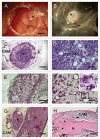
Figure 1
Figure 1
Grafts of human adult kidney progenitor cells, cells from the HeK293 kidney-derived line suspended in Matrigel and grafts of Matrigel only to the chick chorioallantoic membrane (CAM). Suspensions of 2.5 × 10⁶ adult human kidney cells suspended in Matrigel formed compact structures (asterisks) one week after transplantation that apparently attracted host blood vessels (arrows). the graft is shown in situ in the egg in (A) surrounded by a plastic ring that was used to contain the grafted Matrigel. In (B), the graft is shown after removal from the egg and being flipped “upside down” to show the blood vessels beneath. Histological analysis of two different grafts of adult human kidney progenitors from a single donor. (C) is a low-power micrograph of a section through a graft containing Matrigel (M), large amounts of unorganized cells (asterisk) and distinct tubules. Arrows in (C) denote chick blood vessels. At higher magnification in (D), the cuboidal epithelium of the tubules (tub) is easily observed. Red arrowheads point to bright pink-stained erythrocytes in capillaries and small blood vessels that are present throughout the graft. Micrographs of another graft of hAK cells (E and F) show large numbers of tubules of various sizes. two blood vessels (BV) containing magenta-colored erythrocytes are shown at higher magnification in the inset in (F). Grafts of HeK293 cells and Matrigel without cells. (G) is a micrograph of a section through a mass resulting from the grafting of a suspension of cells from the line HeK293, originally derived from human fetal kidney. Large “tumors” (asterisks) formed from the HeK293 cells in the CAM, but no tubular morphogenesis was observed. Large blood sinuses/vessels containing variable amounts of leukocytes penetrated the graft (arrows). When Matrigel (M) only (not containing cells) was transplanted to the CAM, a mass was present containing many blood vessels (arrowheads) after seven days, but was not invaded by other CAM cells (H). Scale bars: (A and B) 1 mm, (C) 500 µm, (D and F) 50 µm, (E and G) 200 µm, (H) 100 µm.
Figure 2
Figure 2
Immuno- and histochemical analysis of CAM grafts of human kidney-derived cells. In (A) a section through a graft of hAK cells stained with a pan-cytokeratin antibody (green) is shown. This section contains mostly cytokeratin-immunopositive tubules. The CAM mesenchyme is unstained (asterisk). (B–D) are micrographs of a section double immunostained with antibodies to cytokeratins (green) and specific for a human lysosome protein (red). Cells in the few tubules present in this section (arrowheads) are labeled with the cytokeratin antibody (B). The human lysosome staining is present in virtually all cells, confirming their derivation from the graft (C). (D) is a merged image showing the overlap of the cytokeratin and lysosome staining of the tubules and the many human lysosome+ cells that had not organized into epithelia. Sections through grafts of HeK293 kidney-derived cells immunostained for human lysosomes (E, red) and cytokeratins (F, green). All the grafted cells in (E), but not the cells of the host CAM (asterisk) are immunopositive for the human cell marker. By contrast, the HeK293 cells are not labeled with the cytokeratin antibody in (F). The immunopositive structure in (F) is the epithelial surface of the CAM (arrow); the antibody used bound avian cytokeratins. Staining of a section through a graft of hAK cells with an antibody to cell division marker Ki67 (G, red) shows cells in a tubule (arrowhead) and an individual cell that are cycling. By contrast, all the cells in a graft of HeK293 (H) cells are dividing. Immunostaining with an antibody to collagen IV, a major component of the basal lamina, shows extensive expression of this protein in a section through a CAM graft of hAK cells (I), while the chick mesenchyme (asterisk) is unstained. Small patches of collagen IV are present in a graft of HeK293 cells (J). Staining of an hAK graft with Dolichos lectin (K) labeled many tubules (i.e., arrowhead), indicating their distal/collecting tubule phenotype. The inset shows several Dolichos-positive (green) and one negative (arrow) tubules in a section through a human fetal kidney. Some AK cells, apparently not part of a tubule, bind Lotus lectin, indicating differentiation to a proximal tubule phenotype (L). In all images, erythrocytes and granulocytes were autofluorescent in both the green and red fluorescent channels and were therefore removed by the spectral unmixing process (see methods). Nuclei (blue) were stained with Hoechst or propidium iodide. The propidium iodide is shown as blue for consistency between the panels. Bar in (A) represents 50 µm in (A, E, F, H–K) and 25 µm in (B–D, G and L).

References

    1. Pleniceanu O, Harari-Steinberg O, Dekel B. Concise review: Kidney stem/progenitor cells: differentiate, sort out or reprogram? Stem Cells. 2010;28:1649–1660. - PMC - PubMed
    1. Nigam SK, Wu W, Bush KT. Organogenesis forum lecture: In vitro kidney development, tissue engineering and systems biology. Organogenesis. 2008;4:137–143. - PMC - PubMed
    1. Bussolati B, Deambrosis I, Russo S, Deregibus MC, Camussi G. Altered angiogenesis and survival in human tumor-derived endothelial cells. FASEB J. 2003;17:1159–1161. - PubMed
    1. Bussolati B, Bruno S, Grange C, Buttiglieri S, Deregibus MC, Cantino D, et al. Isolation of renal progenitor cells from adult human kidney. Am J Pathol. 2005;166:545–545. - PMC - PubMed
    1. Goodpasture EW. A study of human skin grafted upon the chorioallantois of chick embryos. J Exp Med. 1938;68:891–904. - PMC - PubMed

LinkOut - more resources