Clinical Trial
. 2011 Sep;131(9):1916-26.
doi: 10.1038/jid.2011.134. Epub 2011 May 26.

Keratinocyte apoptosis in epidermal remodeling and clearance of psoriasis induced by UV radiation

Affiliations
Free PMC article
Clinical Trial

Keratinocyte apoptosis in epidermal remodeling and clearance of psoriasis induced by UV radiation

Sophie C Weatherhead et al. J Invest Dermatol. 2011 Sep.
Free PMC article

Abstract

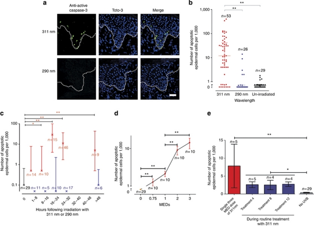
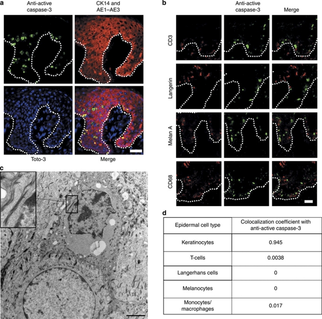
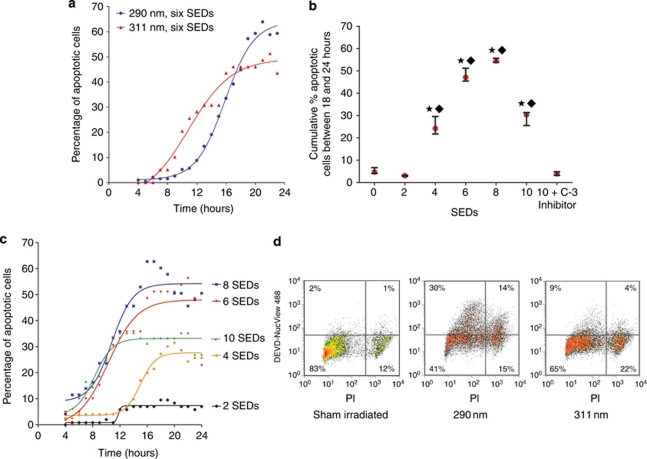
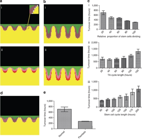
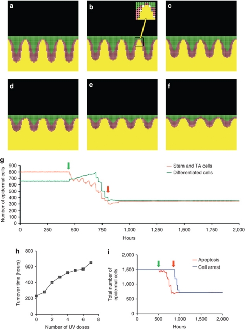
Psoriasis is a common chronic skin disorder, but the mechanisms involved in the resolution and clearance of plaques remain poorly defined. We investigated the mechanism of action of UVB, which is highly effective in clearing psoriasis and inducing remission, and tested the hypothesis that apoptosis is a key mechanism. To distinguish bystander effects, equal erythemal doses of two UVB wavelengths were compared following in vivo irradiation of psoriatic plaques; one is clinically effective (311 nm) and one has no therapeutic effect on psoriasis (290 nm). Only 311 nm UVB induced significant apoptosis in lesional epidermis, and most apoptotic cells were keratinocytes. To determine clinical relevance, we created a computational model of psoriatic epidermis. Modeling predicted apoptosis would occur in both stem and transit-amplifying cells to account for plaque clearance; this was confirmed and quantified experimentally. The median rate of keratinocyte apoptosis from onset to cell death was 20 minutes. These data were fed back into the model and demonstrated that the observed level of keratinocyte apoptosis was sufficient to explain UVB-induced plaque resolution. Our human studies combined with a systems biology approach demonstrate that keratinocyte apoptosis is a key mechanism in psoriatic plaques clearance, providing the basis for future molecular investigation and therapeutic development.


Figures

Figure 1
Figure 1
Significant apoptosis following in vivo irradiation of psoriatic plaques with 311 nm UVB. (a) Representative confocal image of lesional psoriatic epidermis at 18 hours post in vivo irradiation, immunostained with anti-active caspase-3 (green) and Toto-3 (blue). Dotted line shows the basement membrane. Bar=100 μm. (b) Number of anti-active caspase-3 (apoptotic) cells within lesional psoriatic epidermis seen at 16–48 hours following irradiation with either 311 or 290 nm compared with un-irradiated psoriasis. Lines show medians (12/1,000 and 0/1,000 epidermal cells for 311 and 290, respectively) and inter-quartile range. (c) Time-course showing number of apoptotic cells in lesional psoriatic epidermis post UV. Crosses show median number of apoptotic cells per 1,000 epidermal nucleated cells, and inter-quartile range. Red bars represent irradiation with 311 nm, blue represents 290 nm (note median and inter-quartile range is 0 at time-points 0–8, 8–16, and 24–32 hours), and black bar is un-irradiated psoriatic epidermis. (d) Mean (±standard error) number of apoptotic cells in lesional psoriatic epidermis in 10 patients at 24 hours post 311 nm UVB irradiation. Increasing apoptotic response is seen with increasing doses of UVB (means of 1.3, 2.1, 9.1, and 14.3 per 1,000 epidermal cells, respectively), which is significantly different from un-irradiated psoriasis at all doses. (e) Apoptotic response in five patients at 24 hours after a single 311 nm exposure to psoriasis in vivo and at 24 hours after the 4th, 8th, and 12th dose during a routine treatment course. One patient dropped out during treatment because of burning. *P<0.05, **P<0.01. Note the log scale used in figures a, c, and d. MEDs, minimal erythemal doses.
Figure 2
Figure 2
Colocalization of apoptotic cells within lesional psoriatic epidermis with markers specific for epidermal cell types at 24 hours post irradiation with 311 nm UVB. (a, b) Mid Z-section of a confocal image showing colocalization of apoptotic cells (green) with keratinocyte markers (a) and other epidermal cell types (red; b). Bar=100 μm. CD3 is a marker for T-cells, Langerin for Langerhans cells, Melan A for melanocytes, and CD68 for monocyte/macrophages. Dotted line represents the basement membrane. (c) Transmission electron micrograph showing an apoptotic keratinocyte. Insert shows keratin filaments. Desmosomal junctions between the cells were also seen, confirming cells are keratinocytes. Bar=2 μm. (d) Table summarizing the colocalization coefficients for apoptotic cells and the specific epidermal cell markers. Note that a small number of cells (3.4%) could not be identified by colocalization. These cells were at a late stage of apoptosis, when cell surface markers may be lost.
Figure 3
Figure 3
Apoptosis occurs following irradiation with 311 and 290 nm UVB in primary keratinocytes. (a) A membrane-permeable caspase-3 substrate and live cell imaging was used for detection of onset of apoptosis. A greater proportion of apoptotic cells were seen at 24 hours following irradiation with six SEDs of 290 nm than six SEDs of 311 nm, but the time-course was similar. (b) The highest cumulative number of apoptotic keratinocytes occurred following irradiation with six to eight SEDs of 311 nm UVB. The caspase-3-specific inhibitor successfully blocked caspase-3 activity. Median and inter-quartile ranges are shown for three separate experiments. A significant difference in the cumulative proportion of apoptosis was seen between indicated doses and sham-irradiated cells (★) or irradiated cells incubated with caspase-3 and inhibitor (⧫ P<0.05). (c) Time-course of apoptosis following irradiation with increasing doses of 311 nm. Higher doses (six to eight SEDs) were associated with a quicker rate of onset of apoptosis. (d) Flow cytometry showing the effects of irradiation with UVB at a single time point (24 hours). A greater proportion of apoptotic (DEVD-NucView488 caspase-3+) and dead (PI+) cells are observed with 290 nm than 311 nm. PI, propidium iodide; SED, standard erythemal dose.
Figure 4
Figure 4
Development of a mathematical model of normal and psoriatic epidermis based on histological features, differentiation patterns, and epidermal kinetics. (a and bi) Model of normal (a) and psoriatic (b) epidermis with stem cells (white, see insert), actively dividing TA cells (pink), resting TA cells (dark pink), and differentiating cells (green). (a and bii) Two inverted gradients were applied within the model arising from the basement membrane; one maps to the pattern of keratin-10 immunostaining (green) with a specified threshold determining the onset of keratinocyte terminal differentiation; a second gradient contains actively dividing TA cells and stem cells (red), and regulates expansion/contraction of the basement membrane. (c) How adjusting relative proportions of actively dividing stem cells (ci), and TA (cii), or stem cell cycle times (ciii) independently affects turnover time of the whole epidermis. Median and inter-quartile ranges shown; n=50. (d) Model of “normal epidermis” when the number of TA divisions increases from four to five. Note almost all TA cells in the 4th rete ridge are proliferating, but very few in the first, resulting in a patchy distribution of proliferation histologically, not consistent with clinical observation. (e) The turnover times for normal (median 706 hours) and psoriatic (median 270 hours) epidermis used in further experiments. Transit time of differentiated cells (not shown here) reduced from 336 hours to 100 hours.
Figure 5
Figure 5
Mathematical model showing resolution of psoriatic epidermis in response to three MEDs of 311 nm UVB-induced apoptosis. (af) Psoriatic epidermis with elongation of rete ridges and hyperproliferation; different stages of resolution shown until normal state (f) reached; stem cells (white, or gray if apoptosed, see insert), TA cells (pink), and differentiating cells (green). Non-actively dividing stem cells are shown as dark pink. Basement membrane and below is shown in yellow. (g) Progressive reduction in cell numbers following sequential UVB irradiation every 56 hours (equivalent to three times per week). In this experiment, seven doses were required to cause resolution of the psoriasis back to normal. Green arrow shows first irradiation, and red arrow shows final irradiation. (h) Turnover time increases with each dose of UVB as epidermis returns to normal. Irradiation frequency was reduced to 200 hours for the turnover time calculation to allow stability of the model for each measurement. (i) Comparison of time taken for cell numbers to return to normal if apoptosis versus cell cycle arrest is the major mechanism involved in plaque clearance. Note that the time taken for complete resolution following irradiation is significantly prolonged if the mechanism is cell cycle arrest, with a lag time of 336 hours (14 days) from completion of treatment until resolution is completed, although the same number of irradiations were required. Furthermore, in this model remission does not begin until after the UV course is completed, which would not fit with the clinical scenario.

References

    1. Asawanonda P, Anderson RR, Chang Y, et al. 308-nm excimer laser for the treatment of psoriasis: a dose-response study. Arch Dermatol. 2000;136:619–624. - PubMed
    1. Cen H, Mao F, Aronchik I, et al. DEVD-NucView488: a novel class of enzyme substrates for real-time detection of caspase-3 activity in live cells. FASEB J. 2008;22:2243–2252. - PubMed
    1. Coven TR, Burack LH, Gilleaudeau R, et al. Narrowband UV-B produces superior clinical and histopathological resolution of moderate-to-severe psoriasis in patients compared with broadband UV-B. Arch Dermatol. 1997;133:1514–1522. - PubMed
    1. Dawe RS, Wainwright NJ, Cameron H, et al. Narrow-band (TL-01) ultraviolet B phototherapy for chronic plaque psoriasis: three times or five times weekly treatment. Br J Dermatol. 1998;138:833–839. - PubMed
    1. de Cid R, Riveira-Munoz E, Zeeuwen PL, et al. Deletion of the late cornified envelope LCE3B and LCE3C genes as a susceptibility factor for psoriasis. Nat Genet. 2009;41:211–215. - PMC - PubMed

Publication types

MeSH terms