. 2010 Jan 1:3:4-20.
doi: 10.2174/1876536X01003010004.

A Review of Fetal ECG Signal Processing; Issues and Promising Directions

Affiliations

A Review of Fetal ECG Signal Processing; Issues and Promising Directions

Reza Sameni et al. Open Pacing Electrophysiol Ther J. .

Abstract

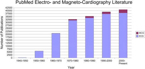
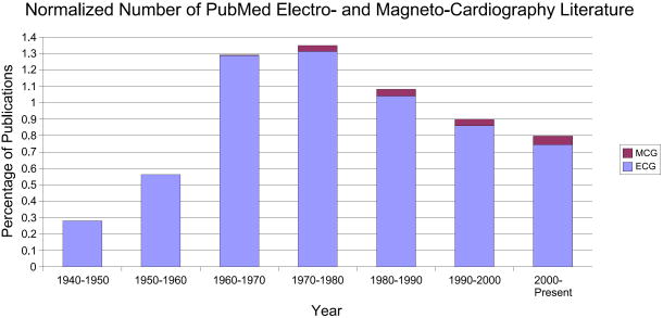
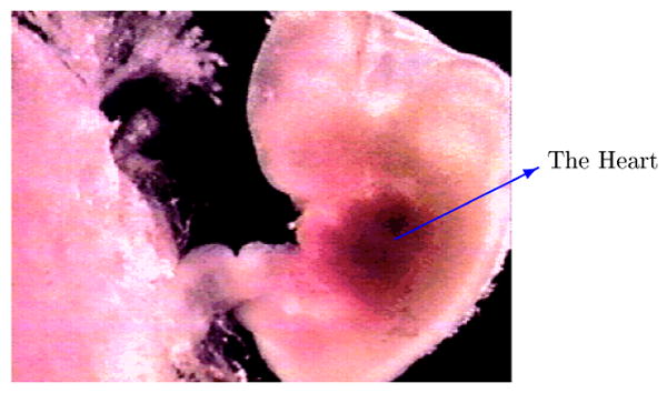
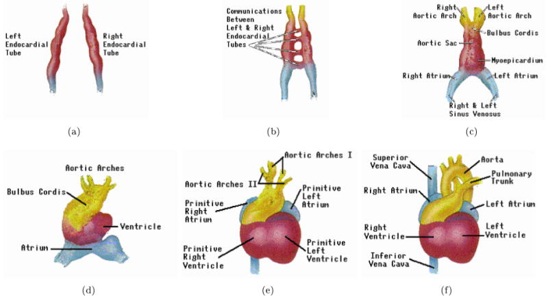
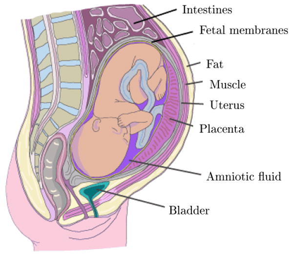
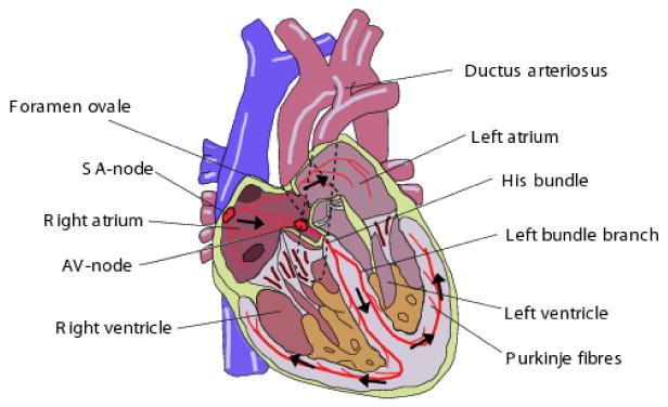
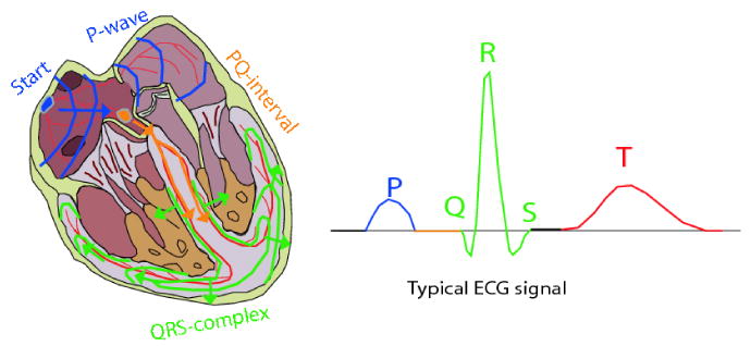
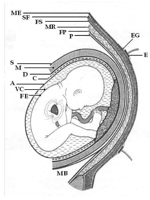
The field of electrocardiography has been in existence for over a century, yet despite significant advances in adult clinical electrocardiography, signal processing techniques and fast digital processors, the analysis of fetal ECGs is still in its infancy. This is, partly due to a lack of availability of gold standard databases, partly due to the relatively low signal-to-noise ratio of the fetal ECG compared to the maternal ECG (caused by the various media between the fetal heart and the measuring electrodes, and the fact that the fetal heart is simply smaller), and in part, due to the less complete clinical knowledge concerning fetal cardiac function and development. In this paper we review a range of promising recording and signal processing techniques for fetal ECG analysis that have been developed over the last forty years, and discuss both their shortcomings and advantages. Before doing so, however, we review fetal cardiac development, and the etiology of the fetal ECG. A selection of relevant models for the fetal/maternal ECG mixture is also discussed. In light of current understanding of the fetal ECG, we then attempt to justify recommendations for promising future directions in signal processing, and database creation.


Conflict of interest statement

Disclosures: Both authors hold shares in MindChild Medical Inc., North Andover, MA, USA and patents in the field of ECG analysis.

Figures

Figure 1
Figure 1
Fetal ECG recorded invasively (upper trace) through a fetal scalp electrode, and non-invasively (lower two traces) through electrodes placed on the mother's abdomen. Fetal heart beats are circled and labeled a, b, c and d.
Figure 2
Figure 2
The amplitude and frequency range of different biosignals, some of which interfere with fetal cardiac signals [9, 10, 11]. The labels in this figure stand for the maternal electrocardiogram (mECG), electroencephalogram (mEEG), electrooculogram (mEOG), electromyogram (mEMG), electrohystrogram (mEHG), and the fetal ECG (fECG). Note that the amplitude of these signals also depends on the site from which the data is recorded.
Figure 3
Figure 3
A general representation of the signal, artifacts and noise present in the ECG in the frequency domain. The region in which the fECG QRS complex manifests is also marked. Note that the fECG largely overlaps with the maternal ECG and other interference. The fECG information also overlaps with the undesired signals in time, space, and feature domains as well; therefore, methods that only work in one specific domain are not able to fully separate the fetal cardiac signals. Image adapted from [12].
Figure 4
Figure 4
Number of publications in the field of fetal electrocardiography and magnetocardiography, registered in PubMed [21].
Figure 5
Figure 5
Number of publications in the field of electrocardiography and magnetocardiography, registered in PubMed [21].
Figure 6
Figure 6
The percentage of publications in the field of electrocardiography and magnetocardiography normalized by the number of publications in all subjects, registered in PubMed [21].
Figure 7
Figure 7
The fetus and its heart in the early stages of development; adapted with permission from [24].
Figure 8
Figure 8
Development stages of the fetal heart during gestation in chronological order (from a to f); adapted with permission from [24].
Figure 9
Figure 9
The major fetomaternal compartments that influence the fetal cardiac surface potentials; adapted with permission from [28]. The vernix caseosa is formed over the fetal skin.
Figure 10
Figure 10
Different fetal presentations and the percentage of incidence at the end of gestation; adapted with permission from [28].
Figure 11
Figure 11
Different fetal vertex positions and their incidence at the end of gestation; adapted with permission from [28]. In the right side abbreviations, L stands for left, R for right, O for occiput, A for anterior, T for transverse and P for posterior. The LOA position is seen in the left graph.
Figure 12
Figure 12
The anatomy of the fetal heart; adapted with permission from [28].
Figure 13
Figure 13
The activation cycle of the fetal heart; adapted with permission from [28].
Figure 14
Figure 14
The increasing complexity in fetal HRV time-series in different gestational ages; adapted with permission from [45].
Figure 15
Figure 15
Upper plot: Mixture of maternal and fetal ECG. Maternal beats are negative spikes (HR = 90 BPM). Fetal beats are the smaller, positive spikes (HR = 138 BPM). Lower plot: Extracted fECG (before denoising). Note that number of beats and relative positions are preserved and so fetal HR estimation and HRV is viable.
Figure 16
Figure 16
Average fetal heart beat morphology taken from scalp electrode (blue line) and average fetal heart beat extracted from abdominal maternal/fetal mixture (magenta line). Note the preservation of clinical features (R-peak, QT-interval, ST-segment).
Figure 17
Figure 17
Schematic representation of the media through which the current generating the fECG is conducted to the surface of the maternal abdomen at approximately 30 weeks. ME: Maternal Epidermis; SF: Subcutaneous Fat; FS: Fascia Superficialis; MR: Musculus Rectus Abdominalis; FP: Fascia Profunda; P: Peritoneum; E: Electrode; EG: Electrode Gel: S: Serosa; M; Myometrium; D: Decidua; C: Chorion; A: Amnion; VC: Vernix Caseosa; FE: Fetal Epidermis; MB; Maternal Bladder. Adapted from [26].

References

    1. Congenital Heart Defects. March of Dimes. 2005. [Online]. Available: http://www.marchofdimes.com/professionals/14332_1212.asp.
    1. Minino AM, Heron MP, Murphy SL, Kochanek KD. Deaths: Final data for 2004. National Vital Statistics Reports. Aug, 2007. - PubMed
    1. Congenital Heart Defects in Children Fact Sheet. American Heart Association; 2008. [Online]. Available: http://www.americanheart.org/children.
    1. Pajkrt E, Weisz B, Firth HV, Chitty LS. Fetal cardiac anomalies and genetic syndromes. Prenat Diagn. Dec, 2004. pp. 1104–1115. [Online]. Available: http://dx.doi.org/10.1002/pd.1067. - DOI - PubMed
    1. Zuckerwar A, Pretlow R, Stoughton J, Baker D. Development of a piezopolymer pressure sensor for a portable fetal heart rate monitor. IEEE Trans Biomed Eng. 1993;40(no. 9):963–969. - PubMed

LinkOut - more resources