

Case Reports
doi: 10.2349/biij.2.2.e33.
Epub 2006 Apr 1.
Phyllodes tumour of the breast
Affiliations
- PMID: 21614232
- PMCID: PMC3097610
- DOI: 10.2349/biij.2.2.e33
Item in Clipboard
Case Reports
Phyllodes tumour of the breast
Biomed Imaging Interv J.
2006 Apr.
No abstract available
Figures

(A) Mediolateral oblique and (B) craniocaudal mammograms show a heterogeneously-dense breast with a round, well-circumscribed, 4.5-cm mass at 3 o’clock in the left breast.

Transverse US image shows a circumscribed, lobulated mass with heterogeneous internal echoes and a slight posterior acoustic enhancement (arrows).

Photograph of an excised specimen shows a well-circumscribed, macrolobulated mass with greyish-white trabeculated cut surface.
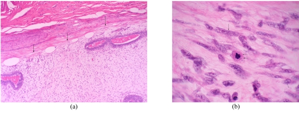
(A) Photomicrograph shows circumscribed border of tumour (arrows) (Haematoxylin & eosin stain, X40). (B) Photomicrograph shows spindle cells with plump nuclei (arrow). Mitosis (double arrows) is also noted. (Haematoxylin & eosin stain, X400).

Benign phyllodes tumour in a 35-year-old woman. (a) Transverse US image shows a circumscribed heterogenous echo with a small cystic space (arrow) and a slight posterior acoustic enhancement. (b) Photomicrograph shows leaf-like processes containing cellular stroma lined with benign ductal epithelial cells projecting into the cystic space (haematoxylin & eosin stain; x100).

Benign phyllodes tumour in a 48-year-old woman. Left craniocaudal mammogram shows a 6-cm lobulated, circumscribed mass in the inner quadrant.
References
-
- Muller J. Uber den feinern bau und die formen der krankhaften geschwulste. Vol. 1. Berlin, Germany: Reimer; 1838. pp. 54–60.
-
- Rosen PP, Oberman HA. Cystosarcoma phyllodes. In: Rosai J, Sobin LH, editors. Atlas of tumor pathology: tumors of the mammary glands. Vol. 7. Wasghington, DC: Armed Forces Institute of Pathology; 1993. pp. 107–14.
-
- Yilmaz E, Sal S, Lebe B. Differentiation of phyllodes tumors versus fibroadenomas. Acta Radiol. 2002;43(1):34–9. - PubMed
-
- Jorge Blanco A, Vargas Serrano B, Rodriguez Romero R, et al. Phyllodes tumors of the breast. Eur Radiol. 1999;9(2):356–60. - PubMed
-
- Czum JM, Sanders LM, Titus JM, et al. Breast imaging case of the day. Benign phyllodes tumor. Radiographics. 1997;17(2):548–51. - PubMed
Publication types
LinkOut - more resources
Full Text Sources