

Glioblastoma multiforme: a rare manifestation of extensive liver and bone metastases
- PMID: 21614314
- PMCID: PMC3097703
- DOI: 10.2349/biij.4.1.e3
Glioblastoma multiforme: a rare manifestation of extensive liver and bone metastases
Abstract
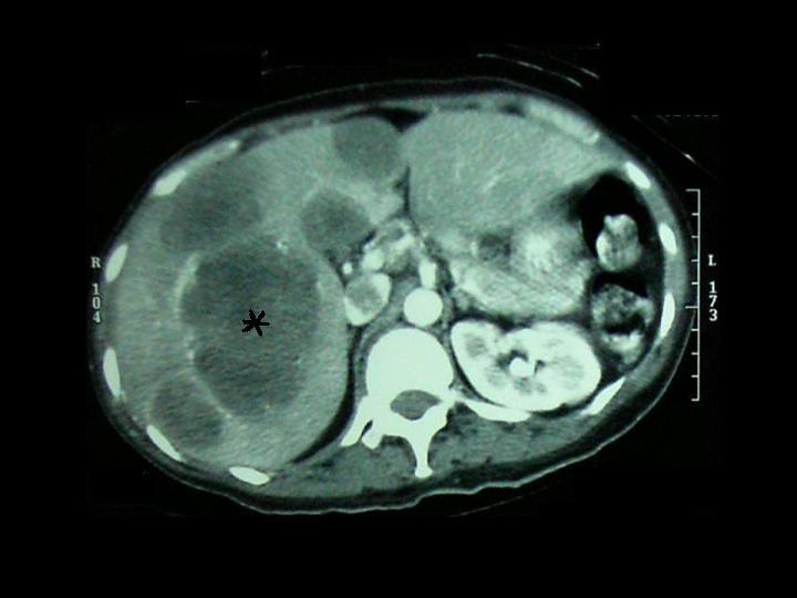
Glioblastoma multiforme (GBM) is the most aggressive form of primary brain tumours known collectively as gliomas. Gliomas are graded by their microscopic appearance. As a rule, their behaviour can be predicted from histology: Grade I (pilocytic astrocytomas) and Grade II (benign astrocytomas) tumours are of low grade and grow slowly over many years. Grade IV tumours (GBM) are the most aggressive and, unfortunately, also the most common in humans, growing rapidly, invading and altering brain function. These tumours arise from the supporting glial cells of the brain during childhood and in adulthood.These growths do not spread throughout the body like other forms of cancer, but cause symptoms by invading the brain. Untreated GBMs are rapidly lethal. Most patients with GBM die of their disease in less than a year and none have long term survival.Extracranial metastases from GBM are extremely rare, with a reported frequency of only 0.44% because of the absence of lymphatics in the brain and the difficulty of tumours to penetrate blood vessels. A case of glioblastoma multiforme with the rare features of extensive liver and bone metastases is presented in this paper.
Keywords: GBM; Glioblastoma multiforme; extracranial metastases; glioma.
Figures


References
-
- Anzil AP. Glioblastoma multiforme with extracranial metastases in the absence of previous craniotomy. Case report. J Neurosurg. 1970;33(1):88–94. - PubMed
-
- Beauchesne P, Soler C, Mosnier JF. Diffuse vertebral body metastasis from a glioblastoma multiforme: a technetium-99m Sestamibi single-photon emission computerized tomography study. J Neurosurg. 2000;93(5):887–90. - PubMed
-
- He J, Olson JJ, James CD. Lack of p16INK4 or retinoblastoma protein (pRb), or amplification-associated overexpression of cdk4 is observed in distinct subsets of malignant glial tumors and cell lines. Cancer Res. 1995;55(21):4833–6. - PubMed
-
- Pasquier B, Pasquier D, N'Golet A, et al. Extraneural metastases of astrocytomas and glioblastomas: clinicopathological study of two cases and review of literature. Cancer. 1980;45(1):112–25. - PubMed
-
- Newton HB, Rosenblum MK, Walker RW. Extraneural metastases of infratentorial glioblastoma multiforme to the peritoneal cavity. Cancer. 1992;69(8):2149–53. - PubMed
Publication types
LinkOut - more resources
Full Text Sources
Other Literature Sources
Research Materials