. 2011 Jul 26;5(7):5729-45.
doi: 10.1021/nn201397z. Epub 2011 Jun 7.

Cell-specific delivery of diverse cargos by bacteriophage MS2 virus-like particles

Affiliations

Cell-specific delivery of diverse cargos by bacteriophage MS2 virus-like particles

Carlee E Ashley et al. ACS Nano. .

Abstract

Virus-like particles (VLPs) of bacteriophage MS2 possess numerous features that make them well-suited for use in targeted delivery of therapeutic and imaging agents. MS2 VLPs can be rapidly produced in large quantities using in vivo or in vitro synthesis techniques. Their capsids can be modified in precise locations via genetic insertion or chemical conjugation, facilitating the multivalent display of targeting ligands. MS2 VLPs also self-assemble in the presence of nucleic acids to specifically encapsidate siRNA and RNA-modified cargos. Here we report the use of MS2 VLPs to selectively deliver nanoparticles, chemotherapeutic drugs, siRNA cocktails, and protein toxins to human hepatocellular carcinoma (HCC). MS2 VLPs modified with a peptide (SP94) that binds HCC exhibit a 10(4)-fold higher avidity for HCC than for hepatocytes, endothelial cells, monocytes, or lymphocytes and can deliver high concentrations of encapsidated cargo to the cytosol of HCC cells. SP94-targeted VLPs loaded with doxorubicin, cisplatin, and 5-fluorouracil selectively kill the HCC cell line, Hep3B, at drug concentrations <1 nM, while SP94-targeted VLPs that encapsidate a siRNA cocktail, which silences expression of cyclin family members, induce growth arrest and apoptosis of Hep3B at siRNA concentrations <150 pM. Impressively, MS2 VLPs, when loaded with ricin toxin A-chain (RTA) and modified to codisplay the SP94 targeting peptide and a histidine-rich fusogenic peptide (H5WYG) that promotes endosomal escape, kill virtually the entire population of Hep3B cells at an RTA concentration of 100 fM without affecting the viability of control cells. Our results demonstrate that MS2 VLPs, because of their tolerance of multivalent peptide display and their ability to specifically encapsidate a variety of chemically disparate cargos, induce selective cytotoxicity of cancer in vitro and represent a significant improvement in the characteristics of VLP-based delivery systems.


Figures
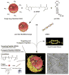
Figure 1
Figure 1. Schematic depicting the process used to synthesize HCC-specific MS2 VLPs that encapsidate chemically disparate therapeutic and imaging agents
Nanoparticles (e.g. quantum dots), protein toxins (e.g. ricin toxin A-chain), and drugs (e.g. doxorubicin) are first conjugated to the pac site using an appropriate crosslinker; for example, quantum dots encapsulated within an amine-terminated PEG layer are linked to a derivative of the pac site that contains a 3′ uracil spacer and sulfhydryl group using the amine-to-sulfhydryl crosslinker, LC-SPDP. Ninety coat protein dimers then self-assemble around RNA-modified cargo to form the 27.5-nm capsid. siRNA molecules drive capsid re-assembly in the absence of the pac site and become incorporated within VLPs at an average concentration of ~85 siRNAs per particle; the yield of fully re-assembled, siRNA-loaded capsids is depicted in the TEM image (scale bar = 50 nm). Cargo-loaded VLPs can be further modified with targeting peptides to promote selective internalization by cancer cells, with fusogenic peptides to promote endosomal escape of internalized VLPs, and with PEG to reduce non-specific interactions and mitigate the humoral immune response against coat protein. Peptides synthesized with a C-terminal cysteine residue are linked to lysine residues (red) on the exterior capsid surface (yellow) via a heterobifunctional crosslinker with a PEG spacer arm.
Figure 2
Figure 2. SP94-targeted MS2 VLPs can selectively deliver disparate therapeutic and imaging agents to human HCC
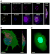
(A) Hyperspectral confocal fluorescence image demonstrating that SP94-targeted VLPs (labeled with Alexa Fluor® 532) can simultaneously deliver ricin toxin A-chain (labeled with Alexa Fluor® 488), quantum dots (Qdot® 585 ITK amino(PEG)), doxorubicin (naturally emits at 560–590 nm), and siRNA (labeled with Alexa Fluor® 647) to Hep3B. Cells were labeled with a blue fluorescent nuclear stain (Hoechst 33342) and a purple fluorescent cytosolic stain (CellTracker Violet BMQC). Scale bars = 20 μm. (B) and (C) Confocal fluorescence microscopy images demonstrating that SP94-targeted VLPs (red) are internalized by Hep3B (B) but not by hepatocytes (C). VLPs were labeled with Alexa Fluor® 555. Cells were labeled with Hoechst 33342 and a green fluorescent cytosolic stain (CellTracker Green CMDFA). Scale bars = 10 μm. VLPs were modified with an average of 60 SP94 peptides/particle and were incubated with cells for 30 minutes at 37°C in all experiments.
Figure 3
Figure 3. SP94-targeted VLPs are directed to lysosomes upon endocytosis by HCC
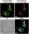
Confocal fluorescence microscopy image demonstrating co-localization between Alexa Fluor® 555-labeled VLPs (red) and an Alexa Fluor® 488-labeled marker for lysosomes (LAMP-1, green) but not between VLPs and an Alexa Fluor® 647-labeled marker for recycling endosomes (Rab11a, white). SlideBook software was used to determine Pearson’s correlation coefficients (r), which are expressed as the mean value ± the standard deviation for n = 3 × 50 cells. Differential Interference Contrast (DIC) images were employed to define the boundaries of Hep3B cells so that pixels outside of the cell boundaries could be disregarded when calculating r-values. VLPs were modified with an average of 60 SP94 peptides/particle and were incubated with cells for 30 minutes at 37°C. Cells were counter-stained with DAPI. Scale bars = 10 μm.
Figure 4
Figure 4. Upon endocytosis, VLPs co-modified with the SP94 targeting peptide and the H5WYG fusogenic peptide become distributed in the cytosol of Hep3B cells, while VLPs modified with just SP94 remain localized in endosomes
(A) – (D) Confocal fluorescence microscopy images of Hep3B cells exposed to SP94-targeted VLPs (red) for either 15 minutes (A and B) or 1 hour (C and D) at 37°C. VLPs were co-modified with ~60 SP94 peptides/particle and ~75 H5WYG peptides/particle in (A) and (C) and with ~60 SP94 peptides/particle alone in (B) and (D). VLPs were labeled with Alexa Fluor® 555. Cells were labeled with Hoechst 33342 and CellTracker Green CMDFA. Scale bars = 10 μm.
Figure 5
Figure 5. SP94-targeted VLPs have a high specific avidity for HCC, and the degree to which they selectively bind to HCC over hepatocytes can be modulated by peptide density and PEGylation
(A) Sample saturation binding curves for SP94-targeted VLPs (~60 peptides/particle) when exposed to Hep3B (Kd = 5.1 × 10¹¹ particles/mL = 0.85 nM) or hepatocytes (Kd = 3.5 × 10¹⁵ particles/mL = 5.8 μM). Saturation binding curves were used to calculate dissociation constants (Kd), which are inversely related to specific avidity. (B) Kd values for VLPs modified with various densities of SP94 or SP88 targeting peptides when exposed to Hep3B. * indicates values are NOT significantly different (using one-way ANOVA, P > 0.05 for n = 5). (C) Kd values for SP94-targeted VLPs (~60 peptides/particle) when exposed to HCC cells (Hep3B, PLC/PRF/5, and HepG2), hepatocytes, endothelial cells (HUVECs), and immune cells (PBMCs and B- and T-lymphocytes). The Kd values of free SP94, VLPs modified with a control peptide that has no known affinity for HCC, and unmodified VLPs (no peptide) when exposed to Hep3B are also given. (D) Kd values for VLPs, modified with the SP94 peptide alone (~60 peptides/particle) or with the SP94 peptide (~60 peptides/particle) and PEG-1000 (~145 molecules/particle) when exposed to Hep3B and hepatocytes. * indicates that values are NOT significantly different (using the unpaired t-test, P > 0.05 for n = 5). Cell concentrations were maintained at 1 × 10⁶ cells/mL in all experiments. All error bars represent 95% confidence intervals (1.96 σ) for n = 5.
Figure 6
Figure 6. SP94-targeted VLPs of MS2 and structurally-related bacteriophages (e.g. Qβ) can deliver a sufficient concentration of chemotherapeutic agents to kill drug-resistant HCC without substantially affecting the viability of hepatocytes
(A) The concentrations of doxorubicin (DOX), cisplatin, 5-fluorouracil (5-FU), a drug cocktail (DOX, cisplatin, and 5-FU), DOX-loaded VLPs, and VLPs loaded with the drug cocktail that are necessary to kill 50% of Hep3B with an induced MDR1+ phenotype (LC50 values) within 24 hours at 37°C. MDR1+ Hep3B were exposed to cyclosporin A (CsA) to reverse Pgp-mediated resistance to DOX. (B) The percentage of MDR1+ Hep3B and hepatocytes that remain viable upon continual exposure to 285 nM of free drugs or drug-loaded VLPs for either 24 hours or 7 days at 37°C; 285 nM is the LC50 value of free DOX when exposed to MDR1+ Hep3B. MS2 and Qβ VLPs were modified with SP94 (~60 peptides/particle) and PEG-1000 (~145 molecules/particle), and cell concentrations were maintained at 1 × 10⁶ cells/mL in all experiments. All error bars represent 95% confidence intervals (1.96 σ) for n = 3.
Figure 7
Figure 7. MS2 VLPs are naturally suited for RNA delivery, and modification of the capsid with SP94 enables targeted delivery of siRNA cocktails that silence expression of various cyclins, causing rapid growth arrest and apoptosis of HCC at picomolar concentrations
(A) The percentage of Hep3B and hepatocytes that become apoptotic upon continual exposure to SP94-targeted, siRNA-loaded VLPs for various periods of time at 37°C. VLPs were loaded with a siRNA cocktail that silences expression of cyclin A2, B1, D1, and E1; the total siRNA concentration was maintained at 150 pM. Cells positive for Alexa Fluor® 647-labeled annexin V were considered to be in the early stages of apoptosis, while cells positive for both annexin V and SYTOX® Green, a cell-impermeant nucleic acid stain, were considered to be in the late stages of apoptosis. (B) The percentage of Hep3B cells that become arrested upon continual exposure to SP94-targeted, siRNA-loaded VLPs for various periods of time at 37°C. VLPs were loaded with a siRNA cocktail that silences expression of cyclin A2, D1, and E1; the total siRNA concentration was 150 pM. The number of proliferating cells was determined by immunofluorescence-based detection of BrdU incorporation, and the number of cells arrested in G0/G1 was determined via Hoechst 33342 staining. (C) The dose-dependent decrease in cyclin A2, B1, D1, and E1 protein expression upon continual exposure of Hep3B to various concentrations of SP94-targeted, siRNA-loaded VLPs for 48 hours at 37°C. The dose-dependent decrease in cyclin A2 mRNA is included for comparison. (D) The time-dependent decrease in cyclin A2, B1, D1, and E1 protein expression upon continual exposure of Hep3B to SP94-targeted, siRNA-loaded VLPs ([siRNA] = 150 pM) at 37°C. The time-dependent decrease in cyclin A2 mRNA is included for comparison. For (C) and (D), VLPs were loaded with a single type of siRNA. Cyclin protein concentrations were determined via immunofluorescence, and cyclin A2 mRNA concentrations were determined by real-time PCR. VLPs were modified with SP94 (~60 peptides/particle), H5WYG (~75 peptides/particle) and PEG-1000 (~145 molecules/particle) in all experiments. All error bars represent 95% confidence intervals (1.96 σ) for n = 3.
Figure 8
Figure 8. SP94-targeted VLPs loaded with cyclin-specific siRNAs selectively transfect HCC with efficiencies similar to that of commercially-available transfection reagents
(A) The concentrations of siRNA necessary to silence 90% of cyclin A2, B1, D1, or E1 protein expression (IC90) in Hep3B when delivered via SP94-targeted VLPs or Lipofectamine RNAiMAX (LFA). (B) The percentage of initial cyclin A2 protein expression that remains upon exposure of Hep3B and hepatocytes to cyclin A2-specific siRNA using SP94-targeted VLPs and LFA, a non-specific transfection reagent, as delivery vehicles. Empty SP94-targeted VLPs, non-targeted, siRNA-loaded VLPs, and siRNA alone were used as controls. VLPs were modified with SP94 (~60 peptides/particle), H5WYG (~75 peptides/particle) and PEG-1000 (~145 molecules/particle) in all experiments. All error bars represent 95% confidence intervals (1.96 σ) for n = 3.
Figure 9
Figure 9. SP94-targeted VLPs that encapsidate ricin toxin A-chain induce apoptosis of HCC at femtomolar concentrations without affecting the viability of hepatocytes
(A) The percentage of Hep3B and hepatocytes that become positive for caspase-3 activation when continually exposed to various concentrations of SP94-targeted, ricin toxin A-chain (RTA)-loaded VLPs for 48 hours at 37°C. (B) The time-dependent activation of caspase-3 in Hep3B and hepatocytes upon exposure to SP94-targeted, RTA-loaded VLPs ([RTA] = 100 fM) at 37°C. Caspase-3 activation was quantified using a FITC-labeled derivative of the caspase-3 inhibitor, DEVD-FMK. (C) and (D) The dose (C) and time (D) dependent decrease in nascent protein synthesis that was observed upon continual exposure of Hep3B to SP94-targeted, RTA-loaded VLPs at 37°C. Cells were exposed to various concentrations of VLPs for 48 hours in (C) and to a fixed concentration of VLPs ([RTA] = 100 fM) for various periods of time in (D). Nascent protein synthesis was quantified using an Alexa Fluor® 488-labeled derivative of methionine. VLPs were modified with SP94 (~60 peptides/particle), H5WYG (~75 peptides/particle), and PEG-1000 (~145 molecules/particle) in all experiments. All error bars represent 95% confidence intervals (1.96 σ) for n = 3.
Figure 10
Figure 10. Co-display of SP94 and H5WYG enables RTA-loaded VLPs to become selectively internalized by HCC and to release their cargo into the cytosol before lysosomal conditions destroy the toxin’s catalytic activity
(A) The percentage of nascent protein synthesis that remains upon exposure of Hep3B and hepatocytes to RTA-loaded VLPs modified with either the SP94 peptide or with a peptide that promotes non-specific macropinocytosis (R8). Empty SP94-modified VLPs, empty R8-modified VLPs, non-targeted VLPs loaded with RTA, and RTA alone were employed as controls. (B) The percentage of Hep3B that become positive for caspase-3 activation when exposed to RTA-loaded VLPs modified with SP94 alone or with a combination of SP94 and H5WYG. Hep3B cells were treated with chloroquine to inhibit lysosomal acidification. Cells were exposed to 100 fM of RTA for 48 hours at 37°C in all experiments. VLPs were modified with PEG-1000 (~145 molecules/particle), as well as an average of ~60 SP94 peptides, ~80 R8 peptides, and/or ~75 H5WYG peptides. All error bars represent 95% confidence intervals (1.96 σ) for n = 3.

References

    1. Peer D, Karp JM, Hong S, FaroKHzad OC, Margalit R, Langer R. Nanocarriers as an Emerging Platform for Cancer Ttherapy. Nature Nanotech. 2007;2:751–760. - PubMed
    1. Peabody DS, Manifold-Wheeler B, Medford A, Jordan SK, do Carmo Caldeira J, Chackerian B. Immunogenic Display of Diverse Peptides on Virus-Like Particles of RNA Phage MS2. J Mol Biol. 2008;380:252–263. - PMC - PubMed
    1. Carrico ZM, Romanini DW, Mehl RA, Francis MB. Oxidative Coupling of Peptides to a Virus Capsid Containing Unnatural Amino Acids. Chem Commun. 2008:1207–1207. - PubMed
    1. Hooker JM, Datta A, Botta M, Raymond KN, Francis MB. Magnetic Resonance Contrast Agents from Viral Capsid Shells: A Comparison of Exterior and Interior Cargo Strategies. Nano Lett. 2007;7:2207–2210. - PubMed
    1. Kovacs EW, Hooker JM, Romanini DW, Holder PG, Berry KE, Francis MB. Dual-Surface-Modified Bacteriophage MS2 as an Ideal Scaffold for a Viral Capsid-Based Drug Delivery System. Bioconjugate Chem. 2007;18:1140–1147. - PubMed

Publication types

MeSH terms