. 2011 Aug;13(8):1200-11.
doi: 10.1111/j.1462-5822.2011.01610.x. Epub 2011 May 25.

The FbaB-type fibronectin-binding protein of Streptococcus pyogenes promotes specific invasion into endothelial cells

Affiliations

The FbaB-type fibronectin-binding protein of Streptococcus pyogenes promotes specific invasion into endothelial cells

Silva Amelung et al. Cell Microbiol. 2011 Aug.

Abstract

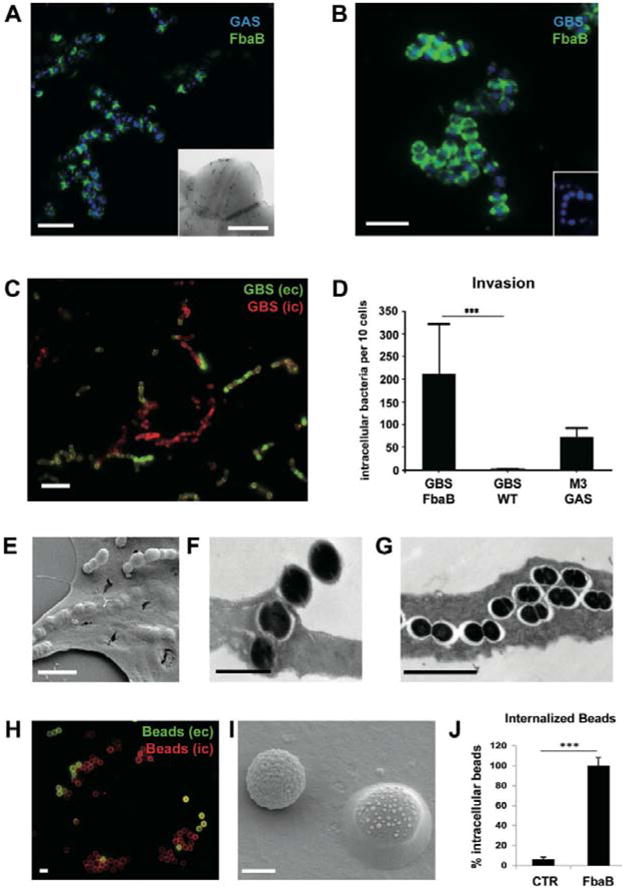
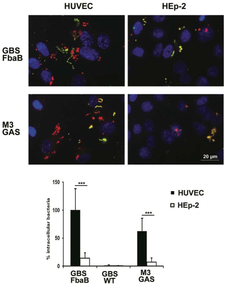
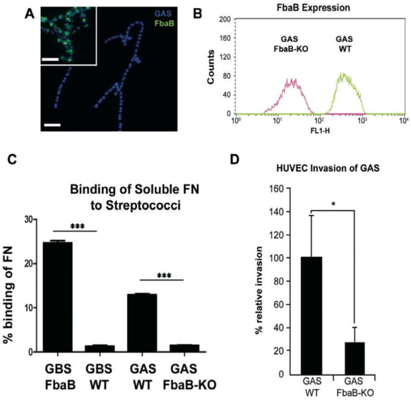
Invasive serotype M3 Streptococcus pyogenes are among the most frequently isolated organisms from patients suffering from invasive streptococcal disease and have the potential to invade primary human endothelial cells (EC) via a rapid and efficient mechanism. FbaB protein, the fibronectin-binding protein expressed by M3 S. pyogenes, was herein identified as a potent invasin for EC. By combining heterologous gene expression with allelic replacement, we demonstrate that FbaB is essential and sufficient to trigger EC invasion via a Rac1-dependent phagocytosis-like uptake. FbaB-mediated uptake follows the classical endocytic pathway with lysosomal destination. FbaB is demonstrated to be a streptococcal invasin exhibiting EC tropism. FbaB thus initiates a process that may contribute to the deep tissue tropism and spread of invasive S. pyogenes isolates into the vascular EC lining.


Figures

Fig. 1
Fig. 1
FbaB mediates invasion into ECs. A. FbaB surface expression of WT M3 GAS. Immunofluorescent (IF)-labelled FbaB (green) on M3 GAS (blue) and FESEM (insert) image of immunolabelled FbaB on M3 GAS. B. Heterologous expression of FbaB on the surface of S. agalactiae (GBS). IF image of immunolabelled FbaB (green) on the surface of the heterologous FbaB-expressing GBS strain (blue), and lack of FbaB expression on WT GBS (insert). C. Invasive potential of GBS-FbaB on EC. HUVEC were infected with GBS-FbaB for 2 h, washed fixed and differentially stained for extra- and intracellular streptococci. IF image of a cell infected with GBS-FbaB; intracellular cocci are red, extracellular green. Figure S1A shows phase contrast of the same field. D. Quantification of EC invasion. Invasion rates were determined by enumerating intracellular (red) bacteria and expressed as intracellular bacteria per 10 cells. The diagram shows invasion rates of GBS-FbaB, the GBS-WT strain and the parental M3 GAS isolate A60 after 2 h of infection (***P < 0.0001). E. FESEM image of adherent and intracellular FbaB-expressing GBS after 2 h of infection. F and G. TEM image of ultrathin sections of GBS-FbaB infected EC after 2 h of infection. H. Uptake of FbaB-coated polystyrene beads into EC. IF image of extracellular (yellow) and intracellular (red) FbaB-coated beads on one EC after 2 h of co-incubation. Figure S1B shows phase contrast of the same field. I. FESEM image of one adherent and one internalized FbaB-coated bead on EC. J. Quantification of bead internalization into HUVEC. Internalization rates were determined after 1 h co-incubation by enumerating intracellular (red) beads. ‘100% Intracellular beads’ refers to a mean of 5.5 FbaB-coated beads per cell (SD ± 0.4). Control beads were coated with GST, revealing a mean of 0.35 intracellular beads per cell (SD ± 0.3; ***P < 0.0002). Bars represent 1 mm (H), 5 mm (A, B, C, E, G), 2 mm (F, I) or 0.5 mm (A, insert).
Fig. 2
Fig. 2
FbaB triggers phagocytosis-like uptake in EC with lysosomal destination. A. Phagocytosis-like uptake of FbaB-expressing GBS into EC. Membrane protrusions along an adherent streptococcal strain characterize the initial uptake process after 30 min of infection. B. Closure of the EC membrane leads to streptococcal internalization. The insert demonstrates formation of equal structures on EC upon internalization of FbaB-coated beads. C. IF image showing F-actin (green) accumulation at the entry site of streptococci (red); the inserts show split channels for F-actin and GBS at higher magnification. D–F. FbaB-mediated uptake follows the classical endocytic pathway. D. IF image of FbaB-GBS (red) that accumulate the early endosomal marker protein EEA1 (green) after EC entry (60 min post infection). The upper insert shows accumulation of the DsRed-labelled PX domain of p40Phox (DsRed-PX, pseudo-coloured in green) around an internalized streptococcal chain (pseudo-coloured in red), the lower insert shows circumferential accumulation of EEA1 (green) on an internalized FbaB-coated bead (red). E. IF image of FbaB-GBS (red) that accumulate the late endosomal/lysosomal marker protein Lamp-1 (green) during the progress of infection (120 min post infection). F. Quantification of inhibition of lysosomal fusion after 2 h of infection using the PI3 kinase (PI3K)-specific inhibitor LY294002. ‘100% Lamp 1 association’ refers to a mean of 22.7 Lamp-1 associated cocci per cell (SD ± 2.4; ***P < 0.002). G. TEM image of ultrathin sections of GBS-FbaB infected EC. Different stages of the fusion events of GBS-FbaB with BSA-gold-loaded terminal lysosomes are shown. The right image shows gold particles in close association with a bacterial cell, indicating successful fusion of the phagosome with a terminal lysosome. Bars indicate 1 mm (A, B, G) and 5 mm (C, D, E).
Fig. 3
Fig. 3
Essential role of the small GTPase Rac1 in the FbaB-mediated EC invasion process. A. Time-lapse microscopy of GBS-FbaB entry into EGFP-WT Rac1-transfected EC. Eight frames of Movie S1 show subsequent accumulation of Rac1 along a streptococcal chain during the first minutes of invasion. B. IF image of GFP-Rac1-expressing EC infected with GBS-FbaB for 60 min (red: F-actin, green: Rac1, blue: FbaB). Inserts at the right side show split channels for F-actin, Rac1 and FbaB stain (from up to down). C. Quantification of FbaB-mediated invasion into EC that express either wild-type Rac1 (WT Rac1), the dominant-negative form of Rac1 (Rac-N17) or EGFP (EGFP) demonstrates that Rac1 is essential for invasion. Invasion rates were determined by enumerating intracellular (red) bacteria after 2 h of infection. EGFP-transfected cells served as control with ‘100% invasion’ referring to 24 intracellular cocci per cell (***P < 0.0001). D. Phase-contrast/fluorescence image of infected EC expressing the PAK-CRIB domain as RFP-fusion protein (CRIB-TagRFP). GBS-FbaB-induced Rac1 activation was visualized by demonstrating accumulation of the PAK-CRIB domain (red) along an invading streptococcal chain after 60 min of infection. The insert shows an enlargement of the fluorescence channel of the indicated area. E. Phase-contrast/fluorescence image of M3 GAS-infected EC expressing the PAK-CRIB domain. The insert shows an enlargement of the fluorescence channel of the indicated area. Bars represent 10 mm (A, B) or 3 mm (D, E).
Fig. 4
Fig. 4
FbaB is an invasin with endothelial cell tropism. HUVEC and HEp-2 cell (human epithelial cell line) were seeded on coverslips, placed into the same well and grown to confluency in EGM2 medium. Cells were infected for 2 h with an moi of 25 with GBS WT or GBS-FbaB, or an moi of 50 with M3 GAS bacteria. Intracellular bacteria were stained in red, extracellular in green. A. IF image of FbaB-expressing GAS and GBS strains infecting HUVEC (left panel) or Hep-2 cells (right panel). Phase-contrast images are shown in Fig. S2A–D. B. Invasion rates were determined by counting intracellular (red) bacteria per cell. ‘100% invasion’ refers to 19 intracellular bacteria per cell [mean of four independent experiments (SD ± 7.3; ***P < 0.0001)].
Fig. 5
Fig. 5
FbaB is the major EC invasin of M3 S. pyogenes. A. Generation of a loss of function mutant (GAS FbaB-KO) in invasive M3 GAS and lack of FbaB surface expression in the deletion mutant. IF image of lacking FbaB surface expression (green) in the mutant, FbaB surface expression of WT M3 GAS (insert in A). GAS were visualized with DAPI (blue). Identical settings were used to acquire both images. Bars represent 5 mm. B. Quantification of FbaB surface expression in the WT M3 GAS strain and the GAS FbaB-KO mutant by flow cytometry analysis. Fluorescence was analysed from 5000 bacteria, using a polyclonal anti-FbaB rabbit antibody in combination with an anti-rabbit Alexa Fluor® 488 secondary antibody. C. FbaB-dependent ‘loss of function’ and ‘gain of function’ of the fibronectin-binding activity on the surface of GAS and GBS. Quantification of fibronectin-binding activity of streptococci either lacking FbaB expression (GBS WT, GAS FbaB-KO) or expressing FbaB (GAS WT, GBS-FbaB) revealed that FbaB is essential but also sufficient to mediate binding of soluble radiolabelled fibronectin in a whole-cell binding assay (***P < 0.0001). D. Quantification of the EC invasion potential of WT GAS in comparison with the GAS FbaB-KO mutant revealed a significant reduction of the invasion potential in the loss of function mutant. Invasion rates were determined after 2 h of infection by counting intracellular (red) bacteria per cell. ‘100% invasion’ refers to 7.4 intracellular bacteria per cell (SD ± 1.9; ***P < 0.0001).

References

    1. Bessen DE, Kalia A. Genomic localization of a T serotype locus to a recombinatorial zone encoding extracellular matrix-binding proteins in Streptococcus pyogenes. Infect Immun. 2002;70:1159–1167. - PMC - PubMed
    1. Catz SD, Johnson JL, Babior BM. The C2A domain of JFC1 binds to 3′-phosphorylated phosphoinositides and directs plasma membrane association in living cells. Proc Natl Acad Sci USA. 2002;99:11652–11657. - PMC - PubMed
    1. Cunningham MW. Pathogenesis of group A streptococcal infections. Clin Microbiol Rev. 2000;13:470–511. - PMC - PubMed
    1. Delvecchio A, Currie BJ, McArthur JD, Walker MJ, Sriprakash KS. Streptococcus pyogenes prtFII, but not sfbI, sfbII or fbp54, is represented more frequently among invasive-disease isolates of tropical Australia. Epidemiol Infect. 2002;128:391–396. - PMC - PubMed
    1. Dinkla K, Rohde M, Jansen WT, Kaplan EL, Chhatwal GS, Talay SR. Rheumatic fever-associated Streptococcus pyogenes isolates aggregate collagen. J Clin Invest. 2003;111:1905–1912. - PMC - PubMed

Publication types

MeSH terms