

Molecular cytogenetic characterization of canine histiocytic sarcoma: A spontaneous model for human histiocytic cancer identifies deletion of tumor suppressor genes and highlights influence of genetic background on tumor behavior
- PMID: 21615919
- PMCID: PMC3121728
- DOI: 10.1186/1471-2407-11-201
Molecular cytogenetic characterization of canine histiocytic sarcoma: A spontaneous model for human histiocytic cancer identifies deletion of tumor suppressor genes and highlights influence of genetic background on tumor behavior
Abstract
Background: Histiocytic malignancies in both humans and dogs are rare and poorly understood. While canine histiocytic sarcoma (HS) is uncommon in the general domestic dog population, there is a strikingly high incidence in a subset of breeds, suggesting heritable predisposition. Molecular cytogenetic profiling of canine HS in these breeds would serve to reveal recurrent DNA copy number aberrations (CNAs) that are breed and/or tumor associated, as well as defining those shared with human HS. This process would identify evolutionarily conserved cytogenetic changes to highlight regions of particular importance to HS biology.
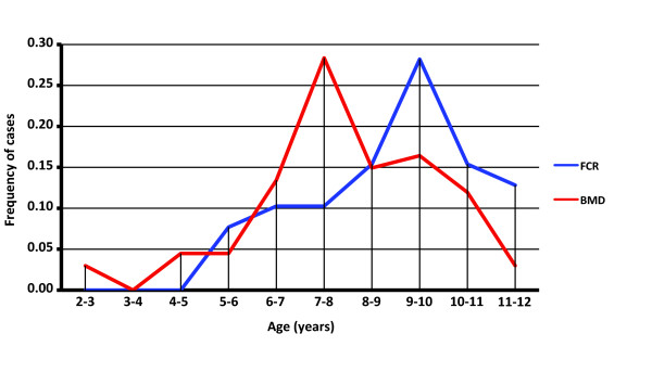
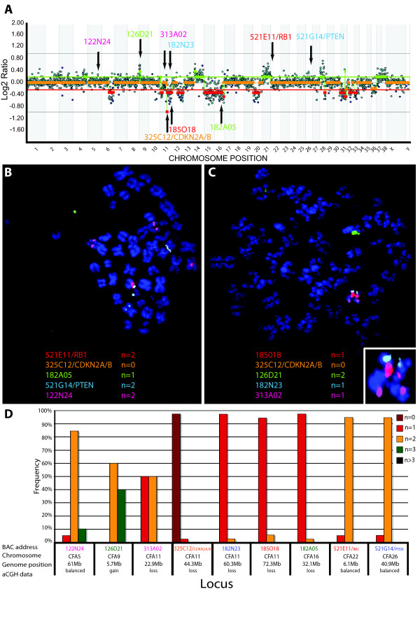
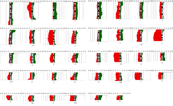
Methods: Using genome wide array comparative genomic hybridization we assessed CNAs in 104 spontaneously occurring HS from two breeds of dog exhibiting a particularly elevated incidence of this tumor, the Bernese Mountain Dog and Flat-Coated Retriever. Recurrent CNAs were evaluated further by multicolor fluorescence in situ hybridization and loss of heterozygosity analyses. Statistical analyses were performed to identify CNAs associated with tumor location and breed.
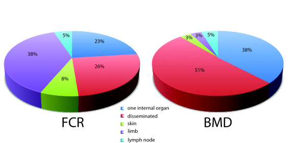
Results: Almost all recurrent CNAs identified in this study were shared between the two breeds, suggesting that they are associated more with the cancer phenotype than with breed. A subset of recurrent genomic imbalances suggested involvement of known cancer associated genes in HS pathogenesis, including deletions of the tumor suppressor genes CDKN2A/B, RB1 and PTEN. A small number of aberrations were unique to each breed, implying that they may contribute to the major differences in tumor location evident in these two breeds. The most highly recurrent canine CNAs revealed in this study are evolutionarily conserved with those reported in human histiocytic proliferations, suggesting that human and dog HS share a conserved pathogenesis.
Conclusions: The breed associated clinical features and DNA copy number aberrations exhibited by canine HS offer a valuable model for the human counterpart, providing additional evidence towards elucidation of the pathophysiological and genetic mechanisms associated with histiocytic malignancies. Extrapolation of data derived from canine histiocytic disorders to human histiocytic proliferation may help to further our understanding of the propagation and cancerization of histiocytic cells, contributing to development of new and effective therapeutic modalities for both species.
Figures
References
-
- Jaffe ES. Pathology and genetics of tumours of haematopeietic tissues. Lyon: World Health Organization of Tumours. International Agency for Research on Cancer; 2001.
-
- Pileri SA, Grogan TM, Harris NL, Banks P, Campo E, Chan JK, Favera RD, Delsol G, De Wolf-Peeters C, Falini B. et al. Tumours of histiocytes and accessory dendritic cells: an immunohistochemical approach to classification from the International Lymphoma Study Group based on 61 cases. Histopathology. 2002;41(1):1–29. doi: 10.1046/j.1365-2559.2002.01418.x. - DOI - PubMed
-
- Favara BE, Feller AC, Pauli M, Jaffe ES, Weiss LM, Arico M, Bucsky P, Egeler RM, Elinder G, Gadner H. et al. Contemporary classification of histiocytic disorders. The WHO Committee On Histiocytic/Reticulum Cell Proliferations. Reclassification Working Group of the Histiocyte Society. Med Pediatr Oncol. 1997;29(3):157–166. doi: 10.1002/(SICI)1096-911X(199709)29:3<157::AID-MPO1>3.0.CO;2-C. - DOI - PubMed
-
- Carrasco DR, Fenton T, Sukhdeo K, Protopopova M, Enos M, You MJ, Di Vizio D, Nogueira C, Stommel J, Pinkus GS. et al. The PTEN and INK4A/ARF tumor suppressors maintain myelolymphoid homeostasis and cooperate to constrain histiocytic sarcoma development in humans. Cancer Cell. 2006;9(5):379–390. doi: 10.1016/j.ccr.2006.03.028. - DOI - PubMed
Publication types
MeSH terms
Substances
LinkOut - more resources
Full Text Sources
Other Literature Sources
Research Materials
Miscellaneous
