. 2011 May 27:11:203.
doi: 10.1186/1471-2407-11-203.

P53 in human melanoma fails to regulate target genes associated with apoptosis and the cell cycle and may contribute to proliferation

Affiliations

P53 in human melanoma fails to regulate target genes associated with apoptosis and the cell cycle and may contribute to proliferation

Kelly A Avery-Kiejda et al. BMC Cancer. .

Abstract

Background: Metastatic melanoma represents a major clinical problem. Its incidence continues to rise in western countries and there are currently no curative treatments. While mutation of the P53 tumour suppressor gene is a common feature of many types of cancer, mutational inactivation of P53 in melanoma is uncommon; however, its function often appears abnormal.

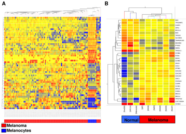
Methods: In this study whole genome bead arrays were used to examine the transcript expression of P53 target genes in extracts from 82 melanoma metastases and 6 melanoma cell lines, to provide a global assessment of aberrant P53 function. The expression of these genes was also examined in extracts derived from diploid human melanocytes and fibroblasts.

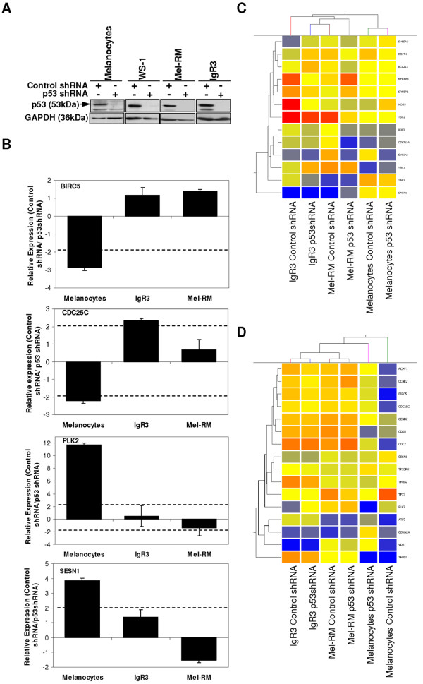
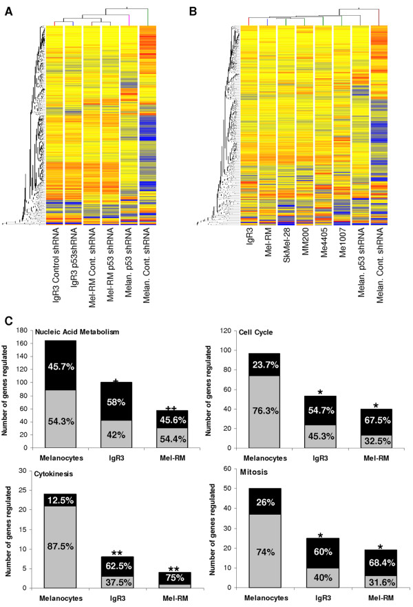
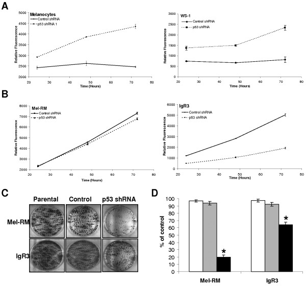
Results: The results indicated that P53 target transcripts involved in apoptosis were under-expressed in melanoma metastases and melanoma cell lines, while those involved in the cell cycle were over-expressed in melanoma cell lines. There was little difference in the transcript expression of P53 target genes between cell lines with null/mutant P53 compared to those with wild-type P53, suggesting that altered expression in melanoma was not related to P53 status. Similarly, down-regulation of P53 by short-hairpin RNA (shRNA) had limited effect on P53 target gene expression in melanoma cells, whereas there were a large number of P53 target genes whose mRNA expression was significantly altered by P53 inhibition in melanocytes. Analysis of whole genome gene expression profiles indicated that the ability of P53 to regulate genes involved in the cell cycle was significantly reduced in melanoma cells. Moreover, inhibition of P53 in melanocytes induced changes in gene expression profiles that were characteristic of melanoma cells and resulted in increased proliferation. Conversely, knockdown of P53 in melanoma cells resulted in decreased proliferation.

Conclusions: These results indicate that P53 target genes involved in apoptosis and cell cycle regulation are aberrantly expressed in melanoma and that this aberrant functional activity of P53 may contribute to the proliferation of melanoma.


Figures

Figure 1
Figure 1
The mRNA expression of P53 target genes is de-regulated in human melanoma. Supervised hierarchical cluster analysis was performed on P53 target genes significantly altered between melanoma and melanocytes. Similarity in the mRNA expression patterns between genes and between samples was measured using Manhattan distance. Distances between clusters represent the average distances between genes and samples in the cluster. Genes are coloured according to their expression level, where up-regulated expression is represented by red, down-regulated expression is represented by blue, and equal expression is represented by yellow. (A) Analysis of 56 differentially expressed genes in 82 metastatic melanoma patients compared to 8 melanocyte cell lines. (B) Analysis of 34 differentially expressed genes in 6 melanoma cell lines (IgR3, Mel-RM, Me1007, MM200, Sk-Mel-28, Me4405) compared to normal cells (melanocytes, FLOW2000, HDF1314).
Figure 2
Figure 2
Inhibition of P53 expression by shRNA alters regulation of P53 target genes. (A) Protein (25 μg) from melanocytes, WS-1, Mel-RM and IgR3 cells that had been stably transduced with P53 shRNA or control shRNA was analysed for the expression of P53 by western blotting. The expression of GAPDH was determined to ensure equal loading. Arrowhead indicates expected molecular weight. (B) Relative quantification of BIRC5, CDC25C, PLK2 and SESN1 mRNA by real-time RT-PCR in melanocytes, Mel-RM and IgR3 cells that had been stably transduced with P53 shRNA or control shRNA. Results are shown as the relative normalised expression (target/β-Actin) of the target gene in cells transduced with control shRNA compared to cells transduced with P53 shRNA (2-ΔΔCt). Values represent the mean ± SE. (C) Supervised hierarchical cluster analysis of 13 genes that were regulated by P53 in melanoma cells only and not in melanocytes. Genes are coloured according to their expression level, where up-regulated expression is represented by red, down-regulated expression is represented by blue, and equal expression is represented by yellow. (D) Supervised hierarchical cluster analysis of 16 genes that were regulated by P53 in melanocytes only and not in melanoma cells. Genes are coloured according to their expression level, where up-regulated expression is represented by red, down-regulated expression is represented by blue, and equal expression is represented by yellow.
Figure 3
Figure 3
Ability of P53 to regulate genes involved in cell cycle is significantly reduced in melanoma. (A) Supervised hierarchical cluster analysis of 728 genes that were significantly regulated by P53 in melanocytes. The relative mRNA expression of these genes in melanocytes, Mel-RM and IgR3 cells that had been stably transduced with either P53 shRNA or control shRNA is shown. (B) Supervised hierarchical cluster analysis of 728 genes that were significantly regulated by P53 in melanocytes. The relative mRNA expression of these genes in melanocytes that had been stably transduced with either P53 shRNA or control shRNA compared to IgR3, Mel-RM, SkMel-28, MM200, Me4405, and Me1007 melanoma cell lines is shown. Genes are coloured according to their expression level, where up-regulated expression is represented by red, down-regulated expression is represented by blue, and equal expression is represented by yellow. (C) The number of genes regulated by P53 (control shRNA versus P53 shRNA) in melanocytes, IgR3 and Mel-RM cell lines in the biological process categories: nucleic acid metabolism, cell cycle, cytokinesis and mitosis as defined by PANTHER [26]. Up-regulated genes are shown in black while down-regulated genes are shown in grey. The number of genes regulated are also depicted as percentages of the total gene list on the bar graph for each of the cell lines. The significance of the regulation of these biological processes by P53 in each of the melanoma cell lines (Mel-RM and IgR3) compared to melanocytes was determined using the gene expression tool in PANTHER (+p < 0.0005, ++p = 0.000003, *p < 0.05, **p < 0.005).
Figure 4
Figure 4
Inhibition of P53 reduces proliferation in melanoma cells. (A) Proliferation was analysed in melanocytes and WS-1 fibroblasts that had been stably transduced with P53 shRNA or control shRNA over a 72 hour period using the MTT assay. Results are represented as the mean ± SE of 3 experiments. (B) Proliferation was analysed in IgR3 and Mel-RM cells that had been stably transduced with P53 shRNA or control shRNA over a 72 hour period using the MTT assay. Results are represented as the mean ± SE of 3 experiments. (C) and (D) Proliferation was analysed by colony formation assay in IgR3 and Mel-RM cells that had been stably transduced with P53 shRNA (black bars) or control shRNA (grey bars) and compared to their parental counterparts (white bars). Representative results are shown in (C) and quantification of 3 independent experiments is shown in (D) with the number of colonies expressed as a percentage of the control shRNA transduced cell lines (mean ± SE). *p < 0.001 by students t-test.

References

    1. Soengas MS, Lowe SW. Apoptosis and melanoma chemoresistance. Oncogene. 2003;22:3138–3151. doi: 10.1038/sj.onc.1206454. - DOI - PubMed
    1. Thompson JF, Scolyer RA, Kefford RF. Cutaneous melanoma. Lancet. 2005;365:687–701. - PubMed
    1. Tsao H, Atkins MB, Sober AJ. Management of cutaneous melanoma. N Engl J Med. 2004;351:998–1012. doi: 10.1056/NEJMra041245. - DOI - PubMed
    1. Soussi T, Beroud C. Assessing TP53 status in human tumours to evaluate clinical outcome. Nat Rev Cancer. 2001;1:233–240. doi: 10.1038/35106009. - DOI - PubMed
    1. Li W, Sanki A, Karim RZ, Thompson JF, Soon Lee C, Zhuang L, McCarthy SW, Scolyer RA. The role of cell cycle regulatory proteins in the pathogenesis of melanoma. Pathology. 2006;38:287–301. doi: 10.1080/00313020600817951. - DOI - PubMed

Publication types

MeSH terms