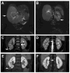
Laparoscopic nephron-sparing resection of synchronous Wilms tumors in a case of hyperplastic perilobar nephroblastomatosis
- PMID: 21616266
- PMCID: PMC3105348
- DOI: 10.1016/j.jpedsurg.2011.01.025
Laparoscopic nephron-sparing resection of synchronous Wilms tumors in a case of hyperplastic perilobar nephroblastomatosis
Abstract
Diffuse hyperplastic perilobar nephroblastomatosis (DHPLN) is a rare precursor lesion of Wilms tumor (WT). Because of the increased risk to develop WT in either kidney, current management algorithms of DHPLN merit nephron-sparing strategies, beginning with chemotherapy and close radiographic monitoring into late childhood. After resolution of DHPLN, subsequent detection of a renal nodule mandates resection to exclude WT. Here, we report the case of a 4-year-old girl who developed 2 synchronous nodules in the right kidney more than 2 years after completion of therapy for DHPLN. Because of the early detection and peripheral location of these 2 nodules, laparoscopic nephron-sparing resection of each was performed using ultrasonic dissection. Both nodules were determined on pathology to be favorable histology WT with negative surgical margins. The child was placed on vincristine and actinomycin D therapy for 18 weeks.
Copyright © 2011 Elsevier Inc. All rights reserved.
Figures


References
-
- Beckwith JB. Nephrogenic rests and the pathogenesis of Wilms tumor: developmental and clinical considerations. Am J Med Genet. 1998 Oct 2;79(4):268–73. - PubMed
-
- Beckwith JB. Precursor lesions of Wilms tumor: clinical and biological implications. Med Pediatr Oncol. 1993;21(3):158–68. - PubMed
-
- Beckwith JB, Kiviat NB, Bonadio JF. Nephrogenic rests, nephroblastomatosis, and the pathogenesis of Wilms' tumor. Pediatr Pathol. 1990;10(1–2):1–36. - PubMed
-
- Perlman EJ, Faria P, Soares A, Hoffer F, Sredni S, Ritchey M, et al. Hyperplastic perilobar nephroblastomatosis: long-term survival of 52 patients. Pediatr Blood Cancer. 2006 Feb;46(2):203–21. - PubMed
-
- Kalapurakal JA, Dome JS, Perlman EJ, Malogolowkin M, Haase GM, Grundy P, et al. Management of Wilms' tumour: current practice and future goals. Lancet Oncol. 2004 Jan;5(1):37–46. - PubMed
Publication types
MeSH terms
Substances
Grants and funding
LinkOut - more resources
Full Text Sources
Medical

