Review
. 2011 Jul;36(7):388-96.
doi: 10.1016/j.tibs.2011.04.005. Epub 2011 May 26.

Mass spectrometry accelerates membrane protein analysis

Affiliations
Review

Mass spectrometry accelerates membrane protein analysis

Jeffrey N Savas et al. Trends Biochem Sci. 2011 Jul.

Abstract

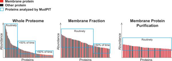
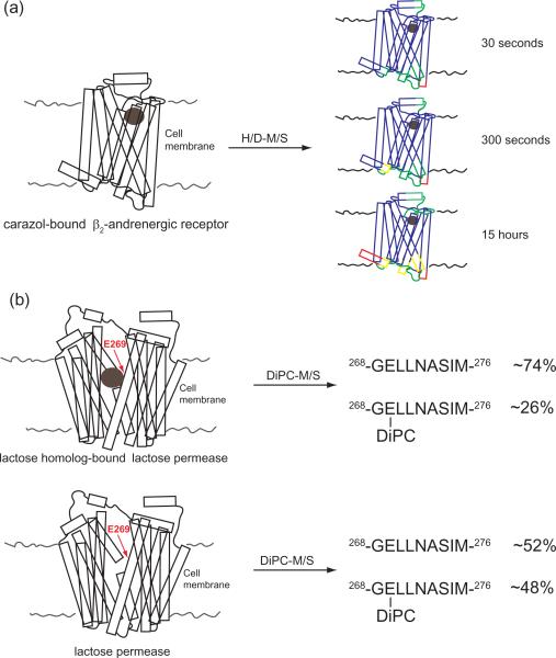
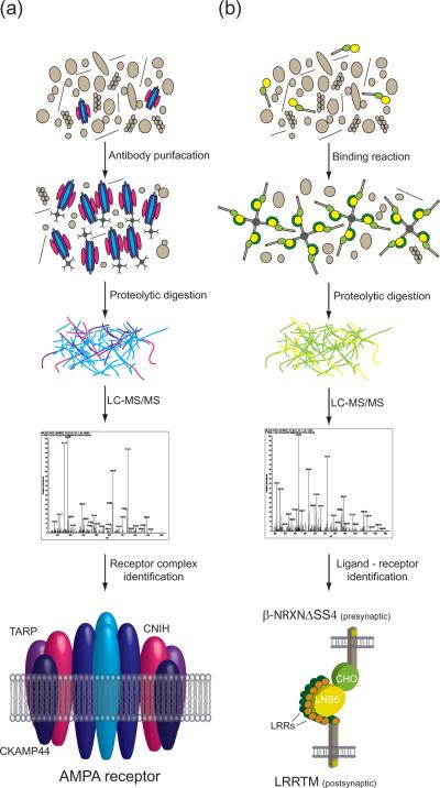
Cellular membranes are composed of proteins and glyco- and phospholipids and play an indispensible role in maintaining cellular integrity and homeostasis, by physically restricting biochemical processes within cells and providing protection. Membrane proteins perform many essential functions, which include operating as transporters, adhesion-anchors, receptors, and enzymes. Recent advancements in proteomic mass spectrometry have resulted in substantial progress towards the determination of the plasma membrane (PM) proteome, resolution of membrane protein topology, establishment of numerous receptor protein complexes, identification of ligand-receptor pairs, and the elucidation of signaling networks originating at the PM. Here, we discuss the recent accelerated success of discovery-based proteomic pipelines for the establishment of a complete membrane proteome.


Figures

Box 1, Figure I
Box 1, Figure I
Views of the membrane proteome based on different enrichment strategies.
Figure 1
Figure 1. Mass spectrometry in conjunction with hydrogen/deuterium (H/D-MS) exchange and carbodiimide diisopropylcarbodiimide (DiPC-MS) chemical probing reveals membrane protein structural characteristics
(a) Upon ligand binding, PM receptors can undergo structural rearrangements. Changes in domain labeling of the ligand-bound GPCR β2-andrenergic receptor (PDB:2RH1), after various exchange durations, is shown as an example of how H/D-MS can be used to probe the molecular details of ligand activation (41). Blue regions indicate protein domains which exchange less than 20%, green regions show 20–60% exchange, yellow 70–80%, and red 90–100%. From these results, one can conclude that some regions of integral membrane proteins are more dynamic than others of this PM protein (41). (b) Specific binding pocket amino acid residues can play an important role in ligand binding of PM receptors. Differential DiPC labeling of lactose permease (PDB:1PV6) in the ligand-bound and ligand-free state revealed a key role for aspartic acid (E)-269 (44). Shown are the ligand-bound (i) and ligand-free (ii) states and the percentage of the 268-GELLNASIM-276 peptide tagged. This analysis revealed a dramatic change in the distribution of E-269 which was modified, from a nearly equal proportion in the free state to a nearly three-fold increase of the unmodified in the bound state.
Figure 2
Figure 2. Membrane protein–protein interactions are identified by biochemical approaches in combination with shotgun proteomics
(a) Antibody purification effectively isolates intact PM receptor complexes from membrane extracts. Purified protein complexes are digested into peptides and subsequently analyzed by LC-MS/MS. The AMPA receptor complex, which has been successfully characterized by antibody purification followed by MS analysis, is shown to illustrate the importance of this approach. AMPA receptor core complexes consist of heterotetramers of glutamate receptor 2 and either glutamate receptor 1, glutamate receptor 3, or glutamate receptor 4 (shown in sky and royal blue). MS-based protein analyses have identified the auxiliary AMPA receptor subunits transmembrane AMPA receptor regulatory proteins (TARPs shown in purple, cornichons (CNIH; fuschia), and cysteine-knot AMPAR modulating protein (CKAMP44, navy blue) (47). (b) In vitro binding reactions with the extracellular domain of receptors as bait and membrane fractions as prey in conjunction with MS can effectively identify ligand–receptor pairs. To illustrate this approach, the ligand–receptor pair (presynaptic neurexin (NRXN) and leucine rich repeat transmembrane protein (LRRTM)) is shown; their interaction was identified by in vitro binding assays followed by MS protein analysis(55). LRRs = Leucine Rich Repeats, CHO = O-linked sugar domain, LNS6 = laminin neurexin and sex hormone-binding protein domain-6.

References

    1. Eichacker LA, et al. Hiding behind hydrophobicity. Transmembrane segments in mass spectrometry. J Biol Chem. 2004;279(49):50915–50922. Translated from eng. in eng. - PubMed
    1. Speers AE, Wu CC. Proteomics of integral membrane proteins--theory and application. Chem Rev. 2007;107(8):3687–3714. Translated from eng. in eng. - PubMed
    1. Hopkins AL, Groom CR. The druggable genome. Nat Rev Drug Discov. 2002;1(9):727–730. Translated from eng. in eng. - PubMed
    1. Weinglass AB, Whitelegge JP, Kaback HR. Integrating mass spectrometry into membrane protein drug discovery. Curr Opin Drug Discov Devel. 2004;7(5):589–599. Translated from eng. in eng. - PubMed
    1. Wu CC, Yates JR., 3rd The application of mass spectrometry to membrane proteomics. Nat Biotechnol. 2003;21(3):262–267. Translated from eng. in eng. - PubMed

Publication types

Substances

LinkOut - more resources