. 2011 Aug 15;184(4):449-58.
doi: 10.1164/rccm.201011-1882OC.

Early alveolar epithelial dysfunction promotes lung inflammation in a mouse model of Hermansky-Pudlak syndrome

Affiliations

Early alveolar epithelial dysfunction promotes lung inflammation in a mouse model of Hermansky-Pudlak syndrome

Elena N Atochina-Vasserman et al. Am J Respir Crit Care Med. .

Abstract

Rationale: The pulmonary phenotype of Hermansky-Pudlak syndrome (HPS) in adults includes foamy alveolar type 2 cells, inflammation, and lung remodeling, but there is no information about ontogeny or early disease mediators.

Objectives: To establish the ontogeny of HPS lung disease in an animal model, examine disease mediators, and relate them to patients with HPS1.

Methods: Mice with mutations in both HPS1/pale ear and HPS2/AP3B1/pearl (EPPE mice) were studied longitudinally. Total lung homogenate, lung tissue sections, and bronchoalveolar lavage (BAL) were examined for phospholipid, collagen, histology, cell counts, chemokines, surfactant protein D (SP-D), and S-nitrosylated SP-D. Isolated alveolar epithelial cells were examined for expression of inflammatory mediators, and chemotaxis assays were used to assess their importance. Pulmonary function test results and BAL from patients with HPS1 and normal volunteers were examined for clinical correlation.

Measurements and main results: EPPE mice develop increased total lung phospholipid, followed by a macrophage-predominant pulmonary inflammation, and lung remodeling including fibrosis. BAL fluid from EPPE animals exhibited early accumulation of both SP-D and S-nitrosylated SP-D. BAL fluid from patients with HPS1 exhibited similar changes in SP-D that correlated inversely with pulmonary function. Alveolar epithelial cells demonstrated expression of both monocyte chemotactic protein (MCP)-1 and inducible nitric oxide synthase in juvenile EPPE mice. Last, BAL from EPPE mice and patients with HPS1 enhanced migration of RAW267.4 cells, which was attenuated by immunodepletion of SP-D and MCP-1.

Conclusions: Inflammation is initiated from the abnormal alveolar epithelial cells in HPS, and S-nitrosylated SP-D plays a significant role in amplifying pulmonary inflammation.


Figures

Figure 1.
Figure 1.
Ontogeny of phospholipid accumulation and inflammation in EPPE mice. Wild-type (WT), EPEP (pale ear), PEPE (pearl), and EPPE (pale ear/pearl) animals were examined at 3 days and 1, 2, 4, 8, 16, and 32 weeks of age. (A) Lung tissue phospholipid (micrograms of phospholipid [PL] per milligram of total protein) from three animals per time point (*P < 0.01 by two-way analysis of variance). (B) Total cell counts from bronchoalveolar lavage (BAL; n = 5–13 animals per time point; *P < 0.01). (C) WT and EPPE lungs were examined after inflation fixation, paraffin sectioning, and hematoxylin and eosin staining (n = 3 animals per age, genotype). Shown is a representative set of photomicrographs from 1- to 32-week WT and EPPE animals at an original magnification of ×20.
Figure 2.
Figure 2.
Ontogeny of pulmonary fibrosis in the pale ear/pearl (EPPE) model of Hermansky-Pudlak syndrome. (A) Wild-type (WT) and EPPE lungs were examined after inflation fixation, paraffin sectioning, and trichrome staining (n = 3 animals per age, genotype). Shown is a representative set of photomicrographs from 32-week WT and EPPE animals at an original magnification of ×10 (top) and ×20 (bottom). The frames in the top panels indicate the images illustrated in the bottom panels. Blue staining indicating collagen deposition is readily seen in the EPPE lung only at 32 weeks of age (arrowhead). (B) Soluble collagen as determined by the Sircol assay in lung tissue from WT and EPPE animals at 16 and 32 weeks of age (n = 7 or 8 animals per group, one-way analysis of variance).
Figure 3.
Figure 3.
EPPE (pale ear/pearl) mice exhibit an increase in bronchoalveolar lavage (BAL) surfactant protein D (SP-D) levels. (A) Equal volumes of BAL from wild-type (WT) and EPPE mice at 2, 4, and 16 weeks of age were analyzed for total SP-D by sodium dodecyl sulfate–polyacrylamide gel electrophoresis, under reduced conditions followed by Western immunoblotting with anti–SP-D antibody. Data shown are representative of three independent experiments (n = 5 in each group). (B) Quantification of SP-D content as described in Methods. Open columns, WT mice; solid columns, EPPE mice. *P < 0.05 versus 2-week WT level; #P < 0.05 versus WT at corresponding age, using one-way analysis of variance.
Figure 4.
Figure 4.
EPPE (pale ear/pearl) animals exhibit S-nitrosylation of surfactant protein D (SP-D) and altered SP-D quaternary structure. (A) Bronchoalveolar lavage (BAL) containing equal amounts of total SP-D (described in the online supplement and shown at bottom) from wild-type (WT) and EPPE mice at various ages was analyzed for S-nitrosylated SP-D (SNO-SP-D) content by the biotin-switch method. Data shown are representative of two separate analyses for each group (n = 5 animals per age, genotype). SNO-SP-D formation consistently increased with age in EPPE animals (A, top). BAL containing equal amounts of total SP-D (A, bottom) was subjected to native gel electrophoresis to determine the quaternary structure of SP-D (A, middle). Data shown are representative of two independent experiments (n = 5 animals per age, genotype). BAL from SP-D–deficient mice (SP-D−/−) was included as a negative control. Multimeric SP-D is incapable of entering the gel at the top, whereas lower molecular weight forms are seen only in BAL from EPPE mice. (B) BAL samples from WT and EPPE mice at 2, 4, and 16 weeks of age were analyzed for total NO metabolites and NO2 as described in Methods, and the data are expressed as the nitrate/nitrite ratio (n = 5; mean ± SEM; *P < 0.05 vs. WT at corresponding age, using one-way analysis of variance).
Figure 5.
Figure 5.
Surfactant protein D (SP-D) modifications and structure disruption occur in human Hermansky-Pudlak syndrome type 1 (HPS1) pulmonary fibrosis and are associated with disease severity. (A) Equal volumes of bronchoalveolar lavage (BAL) fluid from healthy volunteers and patients with HPS1 were analyzed for total SP-D by sodium dodecyl sulfate–polyacrylamide gel electrophoresis, under reduced conditions followed by Western immunoblotting with anti–SP-D antibody. (B) Quantification of total SP-D content. Data are expressed as fold increase compared with normal volunteer samples. Open column, normal volunteers (n = 4); hatched column, patients with HPS1 with minimal to mild lung disease (n = 3); solid column, patients with HPS1 with moderate to severe disease (n = 4). (C) BAL containing equal amounts of total SP-D (described in the online supplement and shown at bottom) were analyzed for S-nitrosylated SP-D (SNO-SP-D) content by the biotin-switch method. Increases in SNO-SP-D formation were associated with increased disease severity (C, top). BAL containing equal amounts of total SP-D was subjected to native gel electrophoresis to determine the quaternary structure of SP-D (C, middle). (D) Quantification of SNO-SP-D content. Data are expressed as the ratio of S-nitrosylated SP-D over total SP-D, and represented as fold change relative to normal volunteers. Open column, normal volunteers (n = 4); hatched column, patients with HPS1 with mild lung disease (n = 3); solid column, patients with HPS1 with severe pulmonary fibrosis (n = 4). *P < 0.05 versus normal volunteers, using one-way analysis of variance.
Figure 6.
Figure 6.
Alveolar epithelial cells (AECs) contribute to early inflammation in animal models of Hermansky-Pudlak syndrome. (A) Bronchoalveolar lavage (BAL) samples were assayed for JE, the mouse homolog of macrophage chemotactic protein-1 (MCP-1), and IL-12p40, using a Searchlight multiplex format (n = 2–6 samples; samples from animals ≤ 2 wk represent pools of 2–4 animals). Data are shown as concentrations (pg/ml) and were analyzed by two-way analysis of variance (ANOVA) (*P < 0.05 vs. wild-type [WT] at corresponding age). (B) Relative real-time polymerase chain reaction (PCR) for MCP-1 and IL-12p40 was performed from AECs or BAL cells isolated from WT and EPPE animals at 2 and 4 weeks of age (ND, none detected; *P < 0.05 vs. WT AECs at corresponding age, **P < 0.01 vs. WT BAL cells at corresponding age by two-way ANOVA). (C) Relative real-time PCR for nitric oxide synthase-2 (NOS2) performed from AECs or BAL cells isolated from WT and EPPE animals at 2 and 4 weeks of age (ND, none detected; *P < 0.05 vs. WT alveolar type 2 [AT2] cells at corresponding age by one-way ANOVA). EPEP = pale ear; EPPE = pale ear/pearl; PEPE = pearl.
Figure 7.
Figure 7.
Alveolar type 2 (AT2) cells from animal models of Hermansky-Pudlak syndrome produce monocyte chemotactic protein-1 (MCP-1). Representative photomicrographs of isolated alveolar epithelial cells from 2-week (A and C) and 4-week (B and D), wild-type (WT) (A and B) and EPPE (pale ear/pearl) (C and D) animals treated with brefeldin for 4 hours (n = 3 experiments each in which AT2 cells were derived from four animals). Samples were examined for the presence of ABCA3 (red; 3c9 antibody) as a marker of AT2 cells and for the presence of MCP-1 (green). 4′,6-Diamidino-2-phenylindole was used to counterstain nuclei (blue).
Figure 8.
Figure 8.
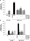
Hermansky-Pudlak syndrome type 1 (HPS1) bronchoalveolar lavage (BAL) induces macrophage chemotaxis in part through S-nitrosylated surfactant protein D (SNO-SP-D). BAL from 4-week-old wild-type (WT) and pale ear/pearl (EPPE) mice was assayed for the ability to induce RAW 264.7 macrophage migration, using a modified Boyden chamber as described in Methods. Briefly, suspensions of RAW 264.7 cells (2 × 10⁶ cells/ml in Dulbecco's modified Eagle's medium) were allowed to migrate toward BAL fluid from 4-week-old WT versus EPPE mice (A), or normal volunteers (NV) versus patients with HPS1 (B) for 3 hours with 5% CO2 at 37°C. Basal migration was expressed as the numbers of cells migrating toward medium only. Positive controls consisted of samples exposed to L-S-nitrosocysteine (L-SNOC) (200 mM) to S-nitrosylate all available proteins from 4-week-old WT (BAL SNO) or NV (NV-SNO). Samples of BAL were also pretreated overnight with anti–SP-D (20 mg/ml) ± anti–MCP-1 (20 mg/ml) antibody followed by immunoprecipitation before adding sample to the Boyden chamber. The importance of SNO-SP-D to chemotaxis was demonstrated by pretreatment of RAW 264.7 cells with anti-CD91 antibody (20 mg/ml) or 20 mM ascorbic acid for 20 minutes before exposure to BAL. Data are normalized as the percentage of cells migrated toward BAL of 4-week EPPE mice (A: *P < 0.05 vs. 4-wk WT mice; #P < 0.05 vs. 4-wk EPPE mice; ζP < 0.05 vs. pretreatment with MCP-1 antibody) or BAL of patients with HPS1 (B: *P < 0.05 vs. NV; #P < 0.05 vs. patients with HPS1). All measurements were performed in triplicate and are representative of three independent experiments analyzed by one-way analysis of variance.

References

    1. Huizing M, Helip-Wooley A, Westbroek W, Gunay-Aygun M, Gahl WA. Disorders of lysosome-related organelle biogenesis: clinical and molecular genetics. Annu Rev Genomics Hum Genet 2008;9:359–386 - PMC - PubMed
    1. Gochuico BR, Huizing M, Golas G, Gahl WA. Interstitial lung disease in Hermansky-Pudlak syndrome-2. Am J Respir Crit Care Med 2010;181:A6018
    1. Nakatani Y, Nakamura N, Sano J, Inayama Y, Kawano N, Yamanaka S, Miyagi Y, Nagashima Y, Ohbayashi C, Mizushima M, et al. Interstitial pneumonia in Hermansky-Pudlak syndrome: significance of florid foamy swelling/degeneration (giant lamellar body degeneration) of type-2 pneumocytes. Virchows Arch 2000;437:304–313 - PubMed
    1. Lyerla TA, Rusiniak ME, Borchers M, Jahreis G, Tan J, Ohtake P, Novak EK, Swank RT. Aberrant lung structure, composition, and function in a murine model of Hermansky-Pudlak syndrome. Am J Physiol Lung Cell Mol Physiol 2003;285:L643–L653 - PubMed
    1. Young LR, Borchers MT, Allen HL, Gibbons RS, McCormack FX. Lung-restricted macrophage activation in the pearl mouse model of Hermansky-Pudlak syndrome. J Immunol 2006;176:4361–4368 - PMC - PubMed

Publication types

MeSH terms