. 2011 Jun;22(6):1064-75.
doi: 10.1681/ASN.2010090956. Epub 2011 May 26.

Suramin inhibits renal fibrosis in chronic kidney disease

Affiliations

Suramin inhibits renal fibrosis in chronic kidney disease

Na Liu et al. J Am Soc Nephrol. 2011 Jun.

Abstract

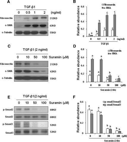
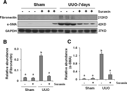
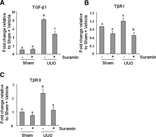
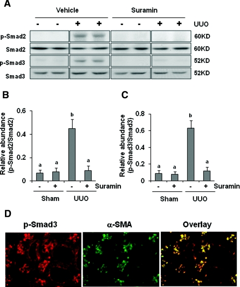
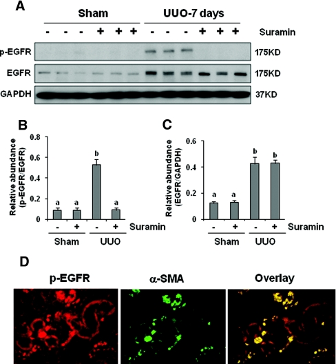
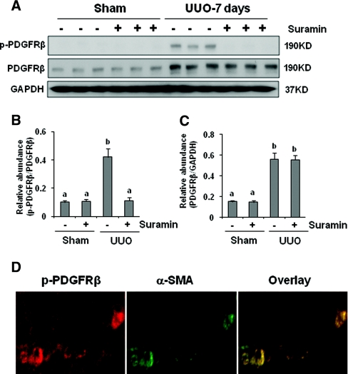
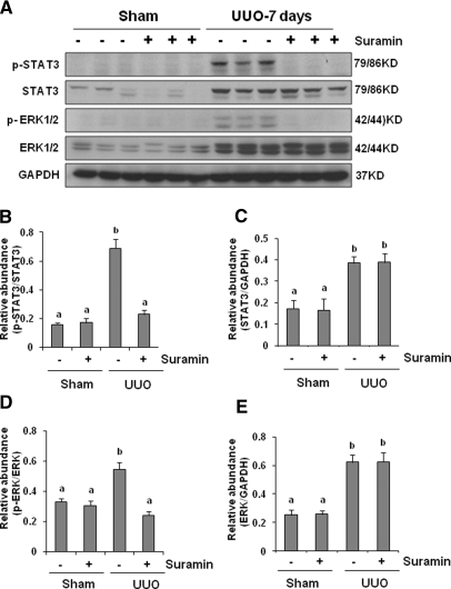
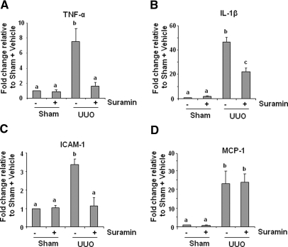
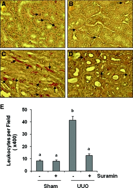
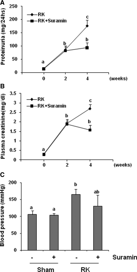
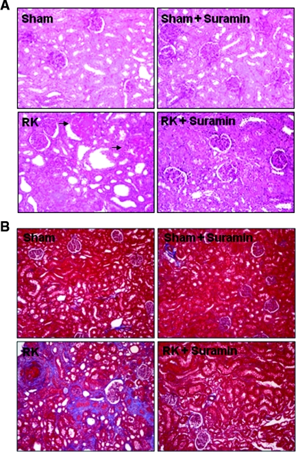
The activation of cytokine and growth factor receptors associates with the development and progression of renal fibrosis. Suramin is a compound that inhibits the interaction of several cytokines and growth factors with their receptors, but whether suramin inhibits the progression of renal fibrosis is unknown. Here, treatment of cultured renal interstitial fibroblasts with suramin inhibited their activation induced by TGF-β1 and serum. In a mouse model of obstructive nephropathy, administration of a single dose of suramin immediately after ureteral obstruction abolished the expression of fibronectin, largely suppressed expression of α-SMA and type I collagen, and reduced the deposition of extracellular matrix proteins. Suramin also decreased the expression of multiple cytokines including TGF-β1 and reduced the interstitial infiltration of leukocytes. Moreover, suramin decreased expression of the type II TGF-β receptor, blocked phosphorylation of the EGF and PDGF receptors, and inactivated several signaling pathways associated with the progression of renal fibrosis. In a rat model of CKD, suramin abrogated proteinuria, limited the decline of renal function, and prevented glomerular and tubulointerstitial damage. Collectively, these findings indicate that suramin is a potent antifibrotic agent that may have therapeutic potential for patients with fibrotic kidney diseases.


Figures

Figure 1.
Figure 1.
Suramin inhibits TGF-β1-induced expression of α-SMA and fibronectin and activation of Smad2/3. Serum-starved NRK-49F cells were incubated with various concentrations of TGF-β1 for (A) 24 hours or (C, E) 24 hours with 2 ng/ml TGF-β1 in the presence of suramin (0 to 100 μM). Cell lysates were prepared and subject to immunoblot analysis with antibodies to α-SMA, fibronectin, phosphorylated Smad2, Smad2, phosphorylated Smad3, Smad3, or α-tubulin. Representative immunoblots from three or more experiments are shown. Expression levels of the indicated proteins were quantified by densitometry and normalized with (B, D) α-tubulin or with total Smad2 or Smad3, respectively. Data are represented as the mean ± SEM. Means with different superscript letters are significantly different from one another (P < 0.05).
Figure 2.
Figure 2.
Suramin blocks on UUO-induced α-SMA and fibronectin expression. (A) Kidney tissue lysates were subjected to immunoblot analysis with specific antibodies against α-SMA, fibronectin, or glyceraldehyde 3-phosphate dehydrogenase (GAPDH). Expression levels of (B) fibronectin and (C) α-SMA were quantified by densitometry and normalized with GAPDH. Data are represented as the mean ± SEM (n = 6). Means with different superscript letters are significantly different from one another (P < 0.05).
Figure 3.
Figure 3.
Suramin attenuates the deposition of ECM and expression of type 1 collagen in obstructed kidneys. (A) Photomicrographs illustrating Masson trichrome staining of kidney tissue after various treatments. (B) Kidney tissue lysates were subject to immunoblot analysis with specific antibodies against collagen I or GAPDH. (C) Expression levels of type 1 collagen were quantified by densitometry and normalized with GAPDH. Data are represented as the mean ± SEM (n = 6). Means with different superscript letters are significantly different from one another (P < 0.05).
Figure 4.
Figure 4.
Suramin suppresses on mRNA expression of TGF-β1 and TGF-β receptors. mRNA was extracted from sham-operated or obstructed kidneys of mice administered with/without suramin and subjected to quantitative real-time RT-PCR as described under Concise Methods. mRNA expression levels of (A) TGF-β1, (B) TβRI, and (C) TβRII were indicated as fold induction over control (sham-operated mice treated with vehicle). Data are represented as the mean ± SEM (n = 6). Means with different superscript letters are significantly different from one another (P < 0.05).
Figure 5.
Figure 5.
Suramin abolishes phosphorylation of Smad2 and Smad3. (A) Kidney tissue lysates were subjected to immunoblot analysis with specific antibodies against Smad2 and Smad3. Expression levels of (B) p-Smad2 and (C) p-Smad3 were quantified by densitometry and normalized with Smad2 and Smad3, respectively. Data are represented as the mean ± SEM (n = 6). Means with different superscript letters are significantly different from one another (P < 0.05). (D) Kidney tissue collected at day 7 was used for co-staining with antibodies to phosphorylated Smad3 and α-SMA.
Figure 6.
Figure 6.
Suramin abolishes phosphorylation of EGFR. Kidney tissue lysates were subjected to immunoblot analysis with specific antibodies against (A) p-EGFR and EGFR. Expression levels of (B) p-EGFR and (C) EGFR were quantified by densitometry and normalized with GAPDH. Data are represented as the mean ± SEM (n = 6). Means with different superscript letters are significantly different from one another (P < 0.05). (D) Kidney tissue collected at day 7 was used for co-staining with antibodies to p-EGFR and α-SMA.
Figure 7.
Figure 7.
Suramin blocks phosphorylation of PDGFR. (A) Kidney tissue lysates were subjected to immunoblot analysis with specific antibodies against (A) phosphorylated PDGF receptor-β (p-PDGFRβ) and PDGFR. Expression levels of (B) p-PDGFRβ and (C) PDGFRβ were quantified by densitometry and normalized with GAPDH. Data are represented as the mean ± SEM (n = 6). Means with different superscript letters are significantly different from one another (P < 0.05). (D) Kidney tissue collected at day 7 was used for co-staining with antibodies to p-PDGFRβ and α-SMA.
Figure 8.
Figure 8.
Suramin blocks phosphorylation of STAT3 and ERK1/2. (A) Kidney tissue lysates were subjected to immunoblot analysis with specific antibodies against phosphorylated STAT3, STAT3, phosphorylated ERK1/2, and ERK1/2. Expression levels of phosphorylated STAT3, STAT3, phosphorylated ERK1/2, and ERK1/2 were quantified by densitometry. Activated (B) STAT3 and (D) ERK1/2 were depicted with phosphorylated STAT3/STAT3 and phosphorylated ERK/ERK ratios, respectively. Protein levels of (C) STAT3 and (E) ERK1/2 were normalized with GADPH. Data are represented as the mean ± SEM (n = 6). Means with different superscript letters are significantly different from one another (P < 0.05).
Figure 9.
Figure 9.
Suramin suppresses on the expression of TNF-α, IL-1β, MCP-1, and ICAM-1. mRNA extracted from kidney tissues were subjected to quantitative real-time RT-PCR as described under Concise Methods. mRNA expression levels of (A) TNF-α, (B) IL-1β, (C) ICAM-1, and (D) MCP-1 were indicated as fold induction over control (sham with vehicle). Data are represented as the mean ± SEM (n = 6). Means with different superscript letters are significantly different from one another (P < 0.05).
Figure 10.
Figure 10.
Suramin inhibits on infiltration of leukocytes. Kidney sections were evaluated for infiltration of neutrophils and monocytes with naphthol AS-D-chloroacetate esterase staining. Photomicrographs illustrate infiltration of neutrophils and monocytes (red color directed by arrows) in different groups: (A) sham with vehicle, (B) sham with suramin, (C) UUO with vehicle, and (D) UUO with suramin. (E) Infiltrated neutrophils and monocytes were counted in three random fields of each sample, and 18 fields (1 × 200) were analyzed for each condition. Data are represented as the mean ± SEM (n = 6). Means with different superscript letters are significantly different from one another (P < 0.05).
Figure 11.
Figure 11.
Suramin improves renal function in 5/6 nephrectomy in rats. Rats received vehicle or suramin from day 14 to week 4 after surgery (n = 10). A parallel study was conducted in rats after sham operation (n = 6). Rats were sacrificed on day 28. (A) Twenty-four-hour urinary protein excretion, (B) serum creatinine, and (C) mean artery pressure. Data are represented as the mean ± SEM. Means with different superscript letters are significantly different from one another (P < 0.05).
Figure 12.
Figure 12.
Surramin attenuates renal histologic damage and ECM protein deposition in 5/6 nephrectomy in rats. (A) Photomicrographs (100×) illustrating periodic-acid–Schiff-stained sections of kidney tissue on week 4 after various treatments as indicated. Sclerotic glomeruli were indicated with arrows. (B) Photomicrographs (100×) illustrating trichrome-stained kidney tissue on week 4 after various treatments. RK, remnant kidney.

References

    1. Wynn TA: Cellular and molecular mechanisms of fibrosis. J Pathol 214: 199–210, 2008 - PMC - PubMed
    1. Neilson EG: Mechanisms of disease: Fibroblasts—A new look at an old problem. Nat Clin Pract Nephrol 2: 101–108, 2006 - PubMed
    1. Lin SL, Kisseleva T, Brenner DA, Duffield JS: Pericytes and perivascular fibroblasts are the primary source of collagen-producing cells in obstructive fibrosis of the kidney. Am J Pathol 173: 1617–1627, 2008 - PMC - PubMed
    1. Boor P, Ostendorf T, Floege J: Renal fibrosis: Novel insights into mechanisms and therapeutic targets. Nat Rev Nephrol 6: 643–656, 2010 - PubMed
    1. Grande MT, Lopez-Novoa JM: Fibroblast activation and myofibroblast generation in obstructive nephropathy. Nat Rev Nephrol 5: 319–328, 2009 - PubMed

Publication types

MeSH terms