. 2011 Jul 8;109(2):193-201.
doi: 10.1161/CIRCRESAHA.111.247023. Epub 2011 May 26.

Interactions between ankyrin-G, Plakophilin-2, and Connexin43 at the cardiac intercalated disc

Affiliations

Interactions between ankyrin-G, Plakophilin-2, and Connexin43 at the cardiac intercalated disc

Priscila Y Sato et al. Circ Res. .

Abstract

Rationale: The early description of the intercalated disc defined 3 structures, all of them involved in cell-cell communication: desmosomes, gap junctions, and adherens junctions. Current evidence demonstrates that molecules not involved in providing a physical continuum between cells also populate the intercalated disc. Key among them is the voltage-gated sodium channel complex. An important component of this complex is the cytoskeletal adaptor protein Ankyrin-G (AnkG).

Objective: To test the hypothesis that AnkG partners with desmosome and gap junction molecules and exerts a functional effect on intercellular communication in the heart.

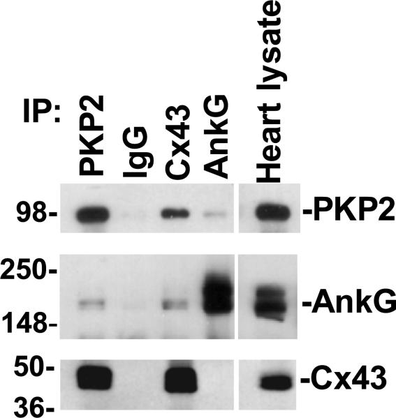
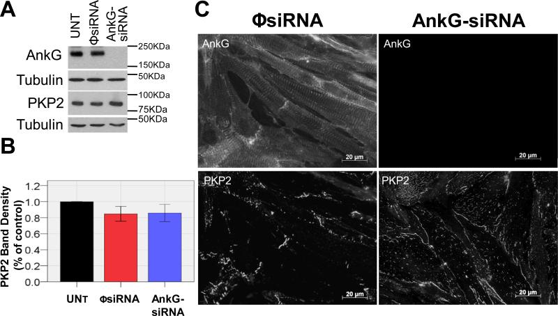
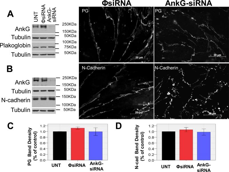
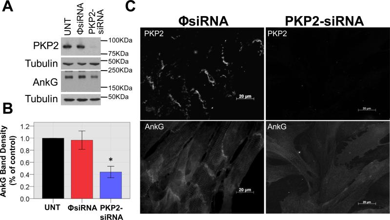
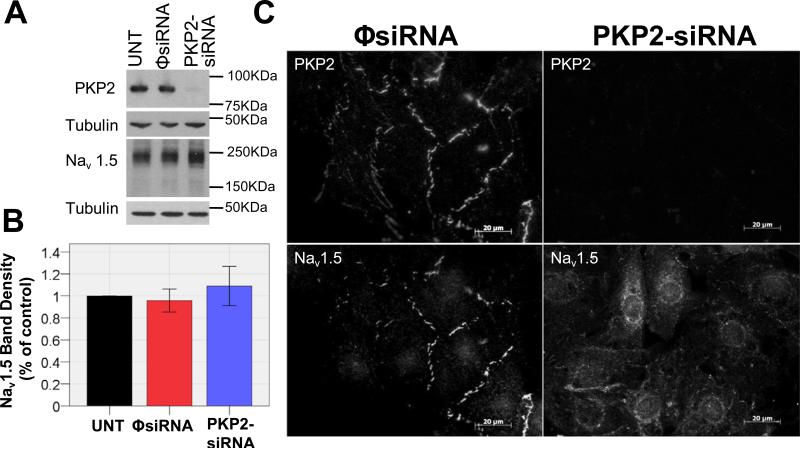
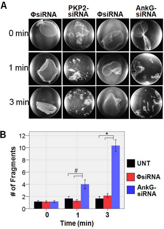
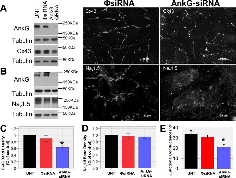
Methods and results: We used a combination of microscopy, immunochemistry, patch-clamp, and optical mapping to assess the interactions between AnkG, Plakophilin-2, and Connexin43. Coimmunoprecipitation studies from rat heart lysate demonstrated associations between the 3 molecules. With the use of siRNA technology, we demonstrated that loss of AnkG expression caused significant changes in subcellular distribution and/or abundance of PKP2 and Connexin43 as well as a decrease in intercellular adhesion strength and electric coupling. Regulation of AnkG and of Na(v)1.5 by Plakophilin-2 was also demonstrated. Finally, optical mapping experiments in AnkG-silenced cells demonstrated a shift in the minimal frequency at which rate-dependence activation block was observed.

Conclusions: These experiments support the hypothesis that AnkG is a key functional component of the intercalated disc at the intersection of 3 complexes often considered independent: the voltage-gated sodium channel, gap junctions, and the cardiac desmosome. Possible implications to the pathophysiology of inherited arrhythmias (such as arrhythmogenic right ventricular cardiomyopathy) are discussed.


Figures

Figure 1
Figure 1
Immunoblots of PKP2, AnkG, and Cx43 (top to bottom) from samples exposed to protein A/G beads coated with PKP2, rabbit IgG (negative control), Cx43, or AnkG antibodies. Heart lysate (far right lane) used as positive control.
Figure 2
Figure 2
Loss of AnkG expression leads to PKP2 relocalization. Panel A: Western blot of NRVMs untreated (UNT) or treated with an oligonucleotide that prevents, or fails to prevent AnkG expression (AnkG-siRNA and ΦsiRNA). Cumulative data in B. All measurements normalized to those from untreated cells in same blot (n=8; p=NS ΦsiRNAvsAnkG-siRNA). C: Immunolocalization of AnkG (top) and PKP2 (bottom) immunoreactive proteins in cells treated with control oligonucleotide (ΦsiRNA) or AnkG-siRNA. Loss of AnkG expression (right column) led to PKP2 redistribution. Bars, 20μm.
Figure 3
Figure 3
Loss of AnkG expression did not affect abundance/distribution of plakoglobin or N-cadherin in NRVMs. A; left: Western blot for AnkG and plakoglobin from same sample; tubulin used for each blot as loading control. Center and right: immunodetection of plakoglobin (PG) in cells treated with control (ΦsiRNA) or AnkG-silencing constructs (AnkG-siRNA). B: Similar conditions. Detection of N-cadherin-immunoreactive proteins. C-D: Quantification of corresponding Western blots. All measurements normalized to those obtained from untreated cells in same blot. In both cases, n=6; p=NS ΦsiRNAvsAnkG-siRNA. Bars, 20μm.
Figure 4
Figure 4
AnkG abundance/distribution in PKP2-silenced NRVMs. A: Western blot for AnkG and PKP2 with respective tubulin loading controls; cells untreated (UNT), treated with siRNA for PKP2 (PKP2-siRNA) or with non-silencing construct (ΦsiRNA). B: Quantification of AnkG band density, corrected individually by loading control and normalized to UNT (n=5; p<0.05 ΦsiRNA vs PKP2-siRNA). C: Immunolocalization of PKP2 (top) and AnkG (bottom) in NRVMs treated with ΦsiRNA or PKP2-siRNA. Bars=20μm.
Figure 5
Figure 5
Loss of PKP2 expression leads to Nav1.5 remodeling. A: Western blot for PKP2 and Nav1.5 with respective tubulin loading controls in NRVMs untreated (UNT), treated with ΦsiRNA, or PKP2-siRNA. B: Quantification of Nav1.5 band density (n=6; pNS; ΦsiRNA vs PKP2-siRNA). C: Immunolocalization of PKP2 (top) and Nav1.5 (bottom) in NRVMs treated with ΦsiRNA or PKP2-siRNA. Bars=20μm.
Figure 6
Figure 6
Loss of AnkG expression weakens intercellular adhesive strength. Cells treated with 1.2-2.4 U/mL dispase 4h to release them from attachment to matrix. A: Pictures of monolayers after dispase treatment (time zero) and after 1 and 3 minutes of gentle shaking (field diameter, 35mm). B: Bars indicate number of fragments found at different times in experiment. An intact, lifted monolayer was counted as 1 fragment; UNT n=6, ΦsiRNA n=7, AnkG-siRNA n=6 (#, p<0.05; *, p<0.01).
Figure 7
Figure 7
Role of AnkG expression on abundance/localization of Cx43 and Nav1.5. A and B: Western blot (left) and immunoreactive signal (center; right) in cells treated with ΦsiRNA (non-silencing control) or AnkG-siRNA. Tubulin used as Western loading control. UNT: Untreated cells. Cells probed for Cx43 or Nav1.5. C: Quantification of Cx43 band density corrected individually by loading controls. Each measurement normalized to that obtained by UNT cells in same blot (p<0.05 AnkG-siRNA vs ΦsiRNA; p<0.01 AnkG-siRNA vs UNT, n=8). D: Quantification of Nav1.5 band density corrected individually by loading controls. Each measurement normalized to UNT cells in same blot. (p=NS, n=5,5,6 for UNT, ΦsiRNA, AnkG-siRNA respectively). E: Dual patch clamp junctional conductance measurements in cell pairs UNT (n=7), treated with ΦsiRNA (n=12) or AnkG-siRNA (n=16). *p<0.01. Bars=20μm.

References

    1. Kaplan SR, Gard JJ, Protonotarios N, Tsatsopoulou A, Spiliopoulou C, Anastasakis A, Squarcioni CP, McKenna WJ, Thiene G, Basso C, Brousse N, Fontaine G, Saffitz JE. Remodeling of myocyte gap junctions in arrhythmogenic right ventricular cardiomyopathy due to a deletion in plakoglobin (Naxos disease). Heart Rhythm. 2004;1:3–11. - PubMed
    1. Musil LS, Cunningham BA, Edelman GM, Goodenough DA. Differential phosphorylation of the gap junction protein connexin43 in junctional communication-competent and -deficient cell lines. J Cell Biol. 1990;111:2077–2088. - PMC - PubMed
    1. Oxford EM, Musa H, Maass K, Coombs W, Taffet SM, Delmar M. Connexin43 remodeling caused by inhibition of plakophilin-2 expression in cardiac cells. Circ Res. 2007;101:703–711. - PubMed
    1. Li J, Patel VV, Kostetskii I, Xiong Y, Chu AF, Jacobson JT, Yu C, Morley GE, Molkentin JD, Radice GL. Cardiac-specific loss of N-cadherin leads to alteration in connexins with conduction slowing and arrhythmogenesis. Circ Res. 2005;97:474–481. - PubMed
    1. Li MW, Mruk DD, Lee WM, Cheng CY. Connexin 43 and plakophilin-2 as a protein complex that regulates blood-testis barrier dynamics. Proc Natl Acad Sci U S A. 2009;106:10213–10218. - PMC - PubMed

Publication types