Review
. 2011 May 27;108(11):1381-91.
doi: 10.1161/CIRCRESAHA.110.234146.

Molecular imaging insights into early inflammatory stages of arterial and aortic valve calcification

Affiliations
Review

Molecular imaging insights into early inflammatory stages of arterial and aortic valve calcification

Sophie E P New et al. Circ Res. .

Abstract

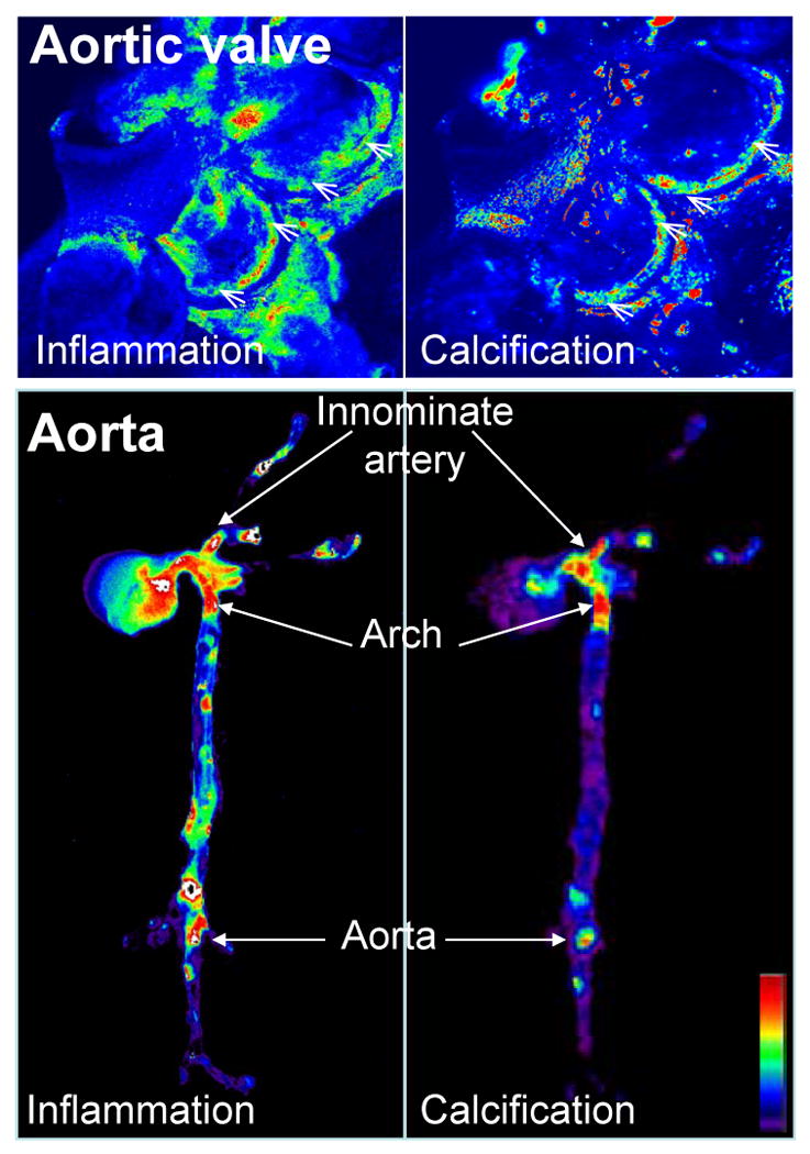
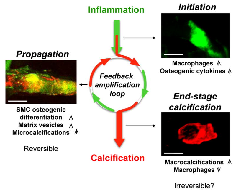
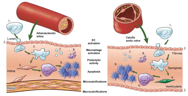
Traditional imaging modalities such as computed tomography, although perfectly adept at identifying and quantifying advanced calcification, cannot detect the early stages of this disorder and offer limited insight into the mechanisms of mineral dysregulation. This review presents optical molecular imaging as a promising tool that simultaneously detects pathobiological processes associated with inflammation and early stages of calcification in vivo at the (sub)cellular levels. Research into treatment of cardiovascular calcification is lacking, as shown by clinical trials that have failed to demonstrate the reduction of calcific aortic stenosis. Hence, the need to elucidate the pathways that contribute to cardiovascular calcification and to develop new therapeutic strategies to prevent or reverse calcification has driven investigations into the use of molecular imaging. This review discusses studies that have used molecular imaging methods to advance knowledge of cardiovascular calcification, focusing in particular on the inflammation-dependent mechanisms of arterial and aortic valve calcification.


Conflict of interest statement

Disclosures: The authors report no conflict of interest.

Figures

Figure 1
Figure 1. Molecular imaging correlated inflammatory activity, defined as macrophage accumulation, with osteogenesis in the aortic valves and aortas of apoE-/- mice
Mice were injected with magneto-fluorescent nanoparticles to visualize macrophage accumulation (left panels), and a spectrally distinct bisphosphonate-imaging agent that binds to nanomolar concentrations of hydroxyapatite to detect osteogenic activity (right panels). In the aortic valve (top) and in the aorta (bottom) molecular imaging detected that inflammatory and osteogenic activities colocalized in the areas of highest mechanical stresses at the aortic valve attachment (arrowheads) and at the atherosclerosis prone areas such as innominate artery, aortic arch, and abdominal aorta (arrows). Images were processed using Osirix software. High signal intensities are shown in red-yellow-green. (Adapted with permission from Aikawa E. et al., Circulation 2009 and Aikawa E. et al., Circulation 2007).
Figure 2
Figure 2. The inflammation-dependent mechanism of calcification visualized by molecular imaging
Sequential intravital fluorescence microscopy was used to observe changes in inflammation and osteogenesis in atherosclerotic arteries. Two spectrally distinct molecular imaging agents were administered through intravenous injection into apoE−/− mice and the common carotid artery was visualized: magneto-fluorescent nanoparticle to target macrophages (green), and a bisphosphonate-imaging agent to detect osteogenic activity/microcalcifications (red). Three different stages could be identified depending on atheroma progression in 20, 30 and 72 week-old apoE-/- mice feed high-fed diet. In the initiation phase – associated with increased macrophages and pro-osteogenic cytokines – only inflammation was observed (green). In the propagation phase – associated with osteogenic activity and generation of microcalcification – inflammation (green) and calcification (red) overlapped (yellow), suggesting that these two processes develop in parallel. Continued inflammation, in parallel with advancing plaque induces further formation of microcalcifications that provoke additional pro-inflammatory responses from macrophages suggesting that feedback amplification loop of calcification and inflammation drives disease progression. Reducing inflammation through anti-inflammatory therapy at this stage may retard osteogenesis and subsequent calcification. In the end-stage phase – associated with increased mineralization and decreased macrophages – macrocalcifications (red) were observed with limited inflammation. Reversing advanced calcification at this late stage is deemed difficult.
Figure 3
Figure 3. Imaging shared molecular mechanisms underlying arterial and aortic valve calcification
The studies cited in this review showed that calcification is an active process composed of a sequence of events, initiated by inflammation and resulting in mineralization. Pro-inflammatory monocytes (1) are recruited to a site via activated endothelial cells (EC) (2). The activation of EC causes increased expression of adhesion molecules, such as VCAM-1, which can be visualized by NIRF imaging using a VCAM-1-targeted agent. Subsequent macrophage (3) accumulation follows, which can be analyzed using NIRF macrophage-targeted nanoparticles. The release of proteolytic enzymes — including matrix metalloproteases and cathepsins — by macrophages, which stimulates the differentiation of myofibroblasts (4) and smooth muscle cells (5) into osteoblasts, can be visualized by molecular imaging using activatable imaging agents. Osteogenic activity in the form of osteoblast (6) formation and microcalcifications associated with generation of calcified matrix vesicles (7) can be identified by bisphosphonate-conjugated imaging agent, while apoptotic bodies (8) can be detected by fluorescently-tagged Annexin A5 imaging probe. Macrocalcifications (9) can be readily detected by molecular imaging and conventional imaging techniques. This schematic visualizes the theory that calcification follows similar processes in both the artery and the aortic valve, and summarizes how NIRF imaging can visualize dynamic sequences of calcification process.

References

    1. Murphy WA, Jr, Nedden Dz D, Gostner P, Knapp R, Recheis W, Seidler H. The iceman: Discovery and imaging. Radiology. 2003;226:614–629. - PubMed
    1. Demer LL, Tintut Y. Mechanisms linking osteoporosis with cardiovascular calcification. Curr Osteoporos Rep. 2009;7:42–46. - PMC - PubMed
    1. Otto CM. Calcific aortic stenosis--time to look more closely at the valve. N Engl J Med. 2008;359:1395–1398. - PubMed
    1. Johnson RC, Leopold JA, Loscalzo J. Vascular calcification: Pathobiological mechanisms and clinical implications. Circ Res. 2006;99:1044–1059. - PubMed
    1. Demer LL, Tintut Y. Vascular calcification: Pathobiology of a multifaceted disease. Circulation. 2008;117:2938–2948. - PMC - PubMed

Publication types