Review
. 2011 May 27;108(11):1392-412.
doi: 10.1161/CIRCRESAHA.110.234138.

Calcific aortic valve stenosis: methods, models, and mechanisms

Affiliations
Review

Calcific aortic valve stenosis: methods, models, and mechanisms

Jordan D Miller et al. Circ Res. .

Abstract

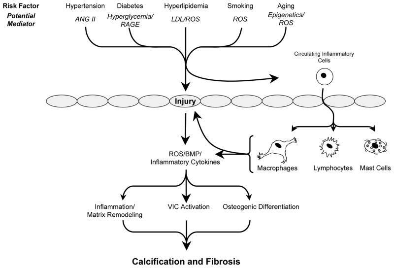
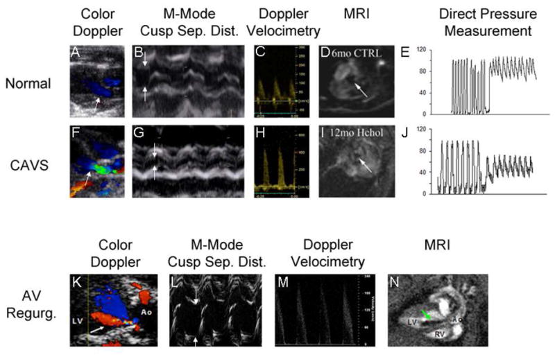
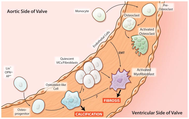
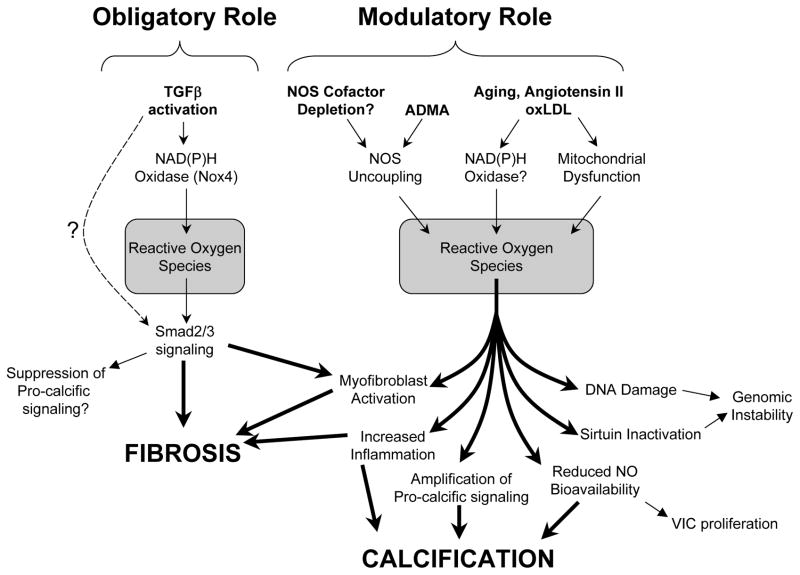
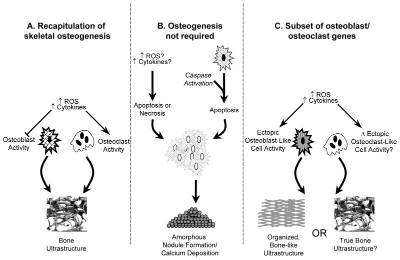
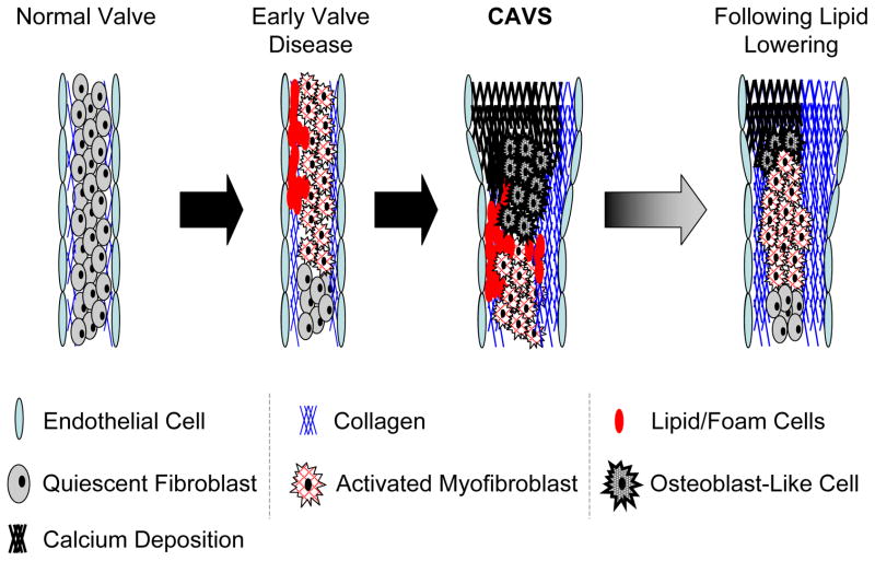
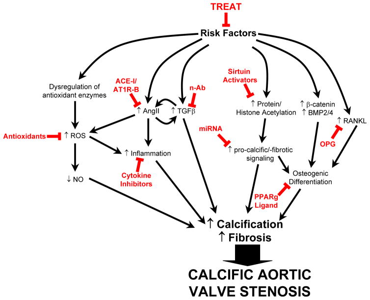
Calcific aortic valve stenosis (CAVS) is a major health problem facing aging societies. The identification of osteoblast-like and osteoclast-like cells in human tissue has led to a major paradigm shift in the field. CAVS was thought to be a passive, degenerative process, whereas now the progression of calcification in CAVS is considered to be actively regulated. Mechanistic studies examining the contributions of true ectopic osteogenesis, nonosseous calcification, and ectopic osteoblast-like cells (that appear to function differently from skeletal osteoblasts) to valvular dysfunction have been facilitated by the development of mouse models of CAVS. Recent studies also suggest that valvular fibrosis, as well as calcification, may play an important role in restricting cusp movement, and CAVS may be more appropriately viewed as a fibrocalcific disease. High-resolution echocardiography and magnetic resonance imaging have emerged as useful tools for testing the efficacy of pharmacological and genetic interventions in vivo. Key studies in humans and animals are reviewed that have shaped current paradigms in the field of CAVS, and suggest promising future areas for research.


Conflict of interest statement

Conflict of Interest

Two of the authors (DDH and RMW) have received osteoprotegerin, and a research grant, from Amgen, Inc.

Figures

Figure 1
Figure 1
Overview of risk factors and potential mechanisms that contribute to calcification and fibrosis of the aortic valve. For clarity, effects of potential mediators on various cell types in the valve have been omitted. Abbreviations: ANG II = angiotensin II; RAGE = receptor for advanced glycosylation end products; LDL = low density lipoproteins; ROS = reactive oxygen species. Grey ovals depict endothelial cells.
Figure 2
Figure 2. Assessment of aortic valve function in mice
Two-dimensional color Doppler images (Panels A,F,K) are used to target M-mode imaging of the aortic valve and Doppler velocimetry, and to assess the presence (arrow, panel K) or absence of aortic regurgitation. Direction of blood flow is indicated by pseudocolors (red = blood flow towards probe, blue = blood flow away from probe). In CAVS, the irregular stenotic valve orifice causes flow acceleration and turbulence (green). The superior spatial and temporal resolution of M-mode echocardiography facilitates quantitation of systolic cusp separation (arrows, panels B,G,L). Doppler velocimetry (Panels C, H, M) allows for estimation of the transvalvular systolic pressure gradient via the Bernoulli Equation. Aortic regurgitation can cause a modest transvalvular systolic pressure gradient even in the absence of reductions in cusp separation (Panel L, green arrow points to regurgitant jet), by virtue of pre-load dependent increases in contractile force and stroke volume and subsequent increases in velocity (Panel M). Magnetic resonance imaging provides temporal and spatial resolution sufficient to portray the aortic valve orifice in two dimensions (arrows, Panels D,I), by virtue of magnetic dephasing of ejected blood. The same principle is employed to depict retrograde flow when aortic regurgitation is present (arrow, Panel N, cine images can be viewed in the Supplemental Movie). Direct pressure measurements can provide incontrovertible evidence of a transvalvular gradient (Panels E and J), but are influenced by the potential for cardiodepression and vasodilation caused by deep general anesthesia, especially in the presence of CAVS. CAVS calcific aortic stenosis; AV Regurg. aortic valve regurgitation; Sep. Dist. separation distance; MRI magnetic resonance imaging.
Figure 3
Figure 3. Potential origins of cells that contribute to valvular calcification and fibrosis
Possible origin of osteoblast-like and osteoclast-like cells in aortic valves in human and murine CAVS. Activated myofibroblasts are likely to come from either quiescent valvular interstitial cells (VICs) or from a sub-population of endothelial cells that undergo endothelial to mesenchymal transformation (EMT). Osteoclast-like cells may originate from circulating monocytes. (Illustration Credit: Cosmocyte/Ben Smith).
Figure 4
Figure 4
Mechanisms whereby reactive oxygen species (ROS) may modulate pro-calcific and pro-fibrotic signaling in CAVS. Nox4-derived ROS may play an obligatory role in TGFβ signaling and induction of fibrosis. In contrast, ROS may play a modulatory role in promoting aortic valve calcification.
Figure 5
Figure 5
Potential pathways contributing to calcified nodule formation in CAVS. A) recapitulation of classical skeletal osteogenesis, in which osteoblast and osteoclast cells respond to exogenous stressors (such as oxidative stress) in a manner similar to that found in bone-derived osteoblasts. B) formation of amorphous calcific nodules without a requirement for osteoblast-like cells, in which stressors initiate cellular aggregation, apoptosis or necrosis, and nodule formation. C) “pseudo-skeletal” ossification, in which cells expressing a subset of osteoblast or osteoclast genes are present in the aortic valve, but respond to exogenous stimuli in fundamentally different ways. For example, previous studies in vitro have shown that—unlike skeletal osteoblasts—cells from cardiovascular tissue typically increase their osteogenic potential in response to exogenous oxidative stress. Bone matrix, replete with marrow hematopoietic elements, has been identified in aortic valves of some patients with CAVS. It is not clear whether this requires processes identical to skeletal osteogenesis (panel A), or if similar structures can be formed by osteoblast-like cells (panel C).
Figure 6
Figure 6
Progression and “regression” of CAVS in “Reversa” mice, . Early stages of CAVS in mice involve myofibroblast activation and lipid insudation/foam cell formation, and are followed by the appearance of osteoblast-like cells, valvular calcification, and substantial increases in valvular fibrosis. Following reduction of blood lipids (“regression” in right panel), there are substantial reductions in valvular lipid content and calcium content, but valvular fibrosis remains increased. Despite reduction of valvular lipid and calcium content, aortic valve function does not improve with substantial lipid lowering.
Figure 7
Figure 7
Potential targets and treatments to slow the progression of aortic valve stenosis. Risk factors (including hypercholesterolemia, hypertension, metabolic syndrome, and smoking) can be treated. Possible signaling cascades and treatments (in red), although supported by some experimental evidence, are speculative. See text for rationale. Abbreviations: ACE-I: angiotensin converting enzyme inhibitor; AT1R-B, angiotension II receptor type I blocker; n-Ab, neutralizing antibody against TGFβ; miRNA, micro-RNA; ROS, reactive oxygen species; NO, nitric oxide; OPG, osteoprotegerin; PPARg, peroxisome proliferator-activated receptor gamma.

References

    1. Nkomo VT, Gardin JM, Skelton TN, Gottdiener JS, Scott CG, Enriquez-Sarano M. Burden of valvular heart diseases: A population-based study. Lancet. 2006;368:1005–1011. - PubMed
    1. Bonow RO, Carabello BA, Chatterjee K, de Leon AC, Jr, Faxon DP, Freed MD, Gaasch WH, Lytle BW, Nishimura RA, O’Gara PT, O’Rourke RA, Otto CM, Shah PM, Shanewise JS. 2008 focused update incorporated into the acc/aha 2006 guidelines for the management of patients with valvular heart disease: A report of the american college of cardiology/american heart association task force on practice guidelines (writing committee to revise the 1998 guidelines for the management of patients with valvular heart disease): Endorsed by the society of cardiovascular anesthesiologists, society for cardiovascular angiography and interventions, and society of thoracic surgeons. Circulation. 2008;118:e523–661. - PubMed
    1. Beckmann E, Grau JB, Sainger R, Poggio P, Ferrari G. Insights into the use of biomarkers in calcific aortic valve disease. J Heart Valve Dis. 2010;19:441–452. - PMC - PubMed
    1. Messika-Zeitoun D, Bielak LF, Peyser PA, Sheedy PF, Turner ST, Nkomo VT, Breen JF, Maalouf J, Scott C, Tajik AJ, Enriquez-Sarano M. Aortic valve calcification: Determinants and progression in the population. Arterioscler Thromb Vasc Biol. 2007;27:642–648. - PubMed
    1. Freeman RV, Otto CM. Spectrum of calcific aortic valve disease: Pathogenesis, disease progression, and treatment strategies. Circulation. 2005;111:3316–3326. - PubMed

Publication types