. 2011 Aug;31(8):1748-56.
doi: 10.1161/ATVBAHA.111.227314. Epub 2011 May 26.

Aging causes collateral rarefaction and increased severity of ischemic injury in multiple tissues

Affiliations

Aging causes collateral rarefaction and increased severity of ischemic injury in multiple tissues

James E Faber et al. Arterioscler Thromb Vasc Biol. 2011 Aug.

Abstract

Objective: Aging is a major risk factor for increased ischemic tissue injury. Whether collateral rarefaction and impaired remodeling contribute to this is unknown. We quantified the number and diameter of native collaterals and their remodeling in 3-, 16-, 24-, and 31-month-old mice.

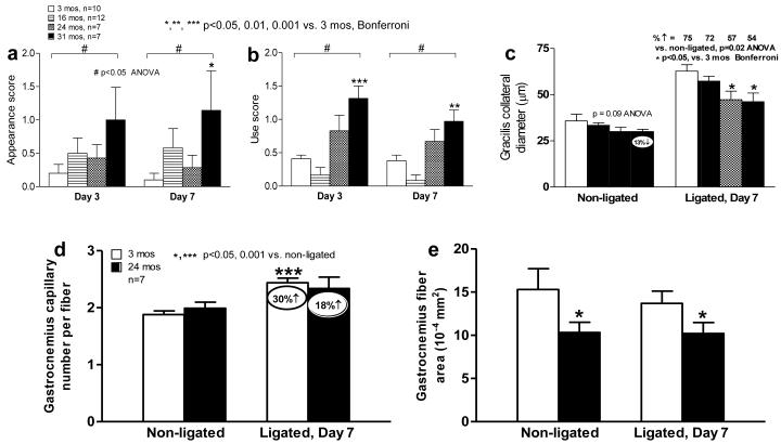
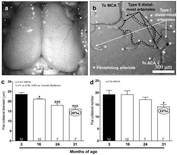
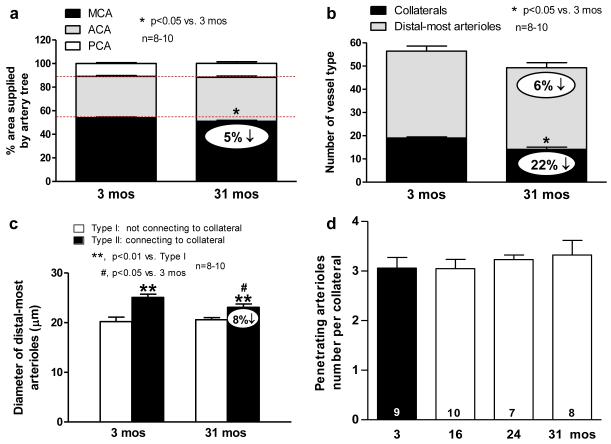
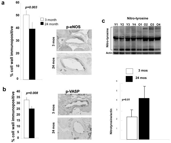
Methods and results: Aging caused an "age-dose-dependent" greater drop in perfusion immediately after femoral artery ligation, followed by a diminished recovery of flow and increase in tissue injury. These effects were associated with a decline in collateral number, diameter, and remodeling. Angiogenesis was also impaired. Mechanistically, these changes were not accompanied by reduced recruitment of T cells or macrophages to remodeling collaterals. However, endothelial nitric oxide synthase signaling was dysfunctional, as indicated by increased protein nitrosylation and less phosphorylated endothelial nitric oxide synthase and vasodilator-stimulated phosphoprotein in collateral wall cells. The cerebral circulation exhibited a similar age-dose-dependent loss of collateral number and diameter and increased tortuosity, resulting in an increase in collateral resistance and infarct volume (eg, 6- and 3-fold, respectively, in 24-month-old mice) after artery occlusion. This was not associated with rarefaction of similarly sized arterioles. Collateral remodeling was also reduced.

Conclusions: Our findings demonstrate that aging causes rarefaction and insufficiency of the collateral circulation in multiple tissues, resulting in more severe ischemic tissue injury.


Figures

Figure 1
Figure 1. Aging causes greater drop in perfusion immediately after femoral artery ligation (FAL), poorer recovery of perfusion, and fewer α-smooth muscle actin (SMA)-positive vessels in adductor collateral zone
a, Laser Doppler perfusion imaging of plantar foot (correlates with leg flow). b, Plantar perfusion. c, Comparison of perfusion immediately after FAL. d, Number of αSMA-positive arterioles in collateral zone of semimembranosis muscle (16 months-old-group missing due to processing complications). Data are mean ± SEM and n-sizes are for number of mice, in this and other figures and tables.
Figure 2
Figure 2. Aging causes collateral circulatory insufficiency in hindlimb
a-c, Greater hindlimb ischemia, use impairment, tendency for decline in native collateral diameter (before FAL), and reduced collateral remodeling after FAL. d, Aging impairs ischemic angiogenesis. e, Baseline muscle atrophy (average muscle fiber cross-sectional area) in aged mice. No ligation-induced atrophy at day-7 after FAL in this moderate ligation model. Decline in baseline collateral diameter and subsequent outward remodeling contribute to findings in Fig 1; less ischemic angiogenesis may also contribute to reduced recovery of perfusion on day-3 and day-7 after FAL.
Figure 3
Figure 3. Aging causes rarefaction of native cerebral collateral diameter and number (a-d)
a,b, Image of dilated, fixed and filled pial cortical circulation; higher magnification image showing two collaterals (black dotted lines) and penetrating arterioles (stars) branching from type II (black arrows) and type I (white arrows) distal-most arterioles that either are or are not cross-connected by collaterals, respectively. MCA, ACA, middle and anterior cerebral artery, respectively. c,d, All collaterals interconnecting MCA and ACA trees in both cerebral hemispheres were quantified. Increased collateral tortuosity and resistance of the native collateral circulation with aging (e-h). e,f, Collateral length (l), axial length of pial collateral. Collateral span (L), scalar length connecting both ends of collateral. h, Relative resistance of the pial collateral circulation, calculated as collateral length / (collateral number x diameter4), is 6- and 10-fold higher in 24- and 31-months-old groups than 3-months-old group. All collaterals between MCA and ACA in both hemispheres were quantified.
Figure 4
Figure 4. Increased cerebral infarct volume and less collateral remodeling in aged mice
a,b, Cerebral infarct volume (A, white area in each slice) was measured 3 days after permanent MCA occlusion. c, Pial collaterals (black arrows) after dilation, fixation and filling with contrast material. d, Native collateral diameter is smaller in aged mice (non-occluded). Aging impairs collateral remodeling (% increase is relative to 3-months-old, p=0.03); inset, change in diameter. White bar, 3-months-old; black bar, 24-months-old.
Figure 5
Figure 5. Cerebral morphometry in young and old mice
a. Percent area of cortex supplied by MCA, ACA and PCA trees. 5% smaller MCA territory with aging; this favors smaller infarct volumes, opposite to Figure 6. b. MCA tree morphometry: similar 6% decrease in distal-most arterioles (DMA, both Type I and II) of MCA tree as 5% decrease in MCA territory. Collateral number decreased by similar absolute amount (though greater percentage) as DMAs, indicating concomitant loss of both. c, Type II DMAs are larger than Type I, consistent with greater flow of Type 2 to supply penetrating arterioles branching from collaterals. Aging decreases Type II diameter. This is consistent with decline in diameter of the collaterals they supply (Figure 3), which may relate to the mechanism of collateral loss (see Text). d, Collaterals not lost have same number of penetrating arterioles branching from them.
Figure 6
Figure 6. Phospho-eNOS and phospho-VASP are decreased in aged collaterals
Immunohistochemistry of gracilis muscle collaterals for phosphorylated eNOS (a) and phosphorylated VASP (b), and for nitrotyrosine in mesenteric arterioles (c) in 3-months-old (Y1-Y4, young) and 24-months-old (O1-O4, old) mice.

References

    1. Cooper LT, Cooke JP, Dzau VJ. The vasculopathy of aging. J Gerontol. 1994;49:B191–196. - PubMed
    1. Lloyd-Jones D, Adams RJ, Brown TM, Carnethon M, Dai S, De Simone G, Ferguson TB, Ford E, Furie K, Gillespie C, Go A, Greenlund K, Haase N, Hailpern S, Ho PM, Howard V, Kissela B, Kittner S, Lackland D, Lisabeth L, Marelli A, McDermott MM, Meigs J, Mozaffarian D, Mussolino M, Nichol G, Roger VL, Rosamond W, Sacco R, Sorlie P, Stafford R, Thom T, Wasserthiel-Smoller S, Wong ND, Wylie-Rosett J. Heart disease and stroke statistics--2010 update: a report from the American Heart Association. Circulation. 2010;121:e46–e215. - PubMed
    1. Pedelty L, Gorelick PB. Management of hypertension and cerebrovascular disease in the elderly. Am J Med. 2008;121(8 Suppl):S23–31. - PubMed
    1. Eliason JL, Wainess RM, Proctor MC, Dimick JB, Cowan JA, Jr, Upchurch GR, Jr, Stanley JC, Henke PK. A national and single institutional experience in the contemporary treatment of acute lower extremity ischemia. Ann Surg. 2003;238:382–90. - PMC - PubMed
    1. Ohta T, Hosaka M, Ishibashi H, Sugimoto I, Mihara E, Hida K, Takeuchi N, Hachiya J, Kato M, Kazui H, Nagata Y. Limb salvage and survival rates among elderly patients with advanced limb ischemia. Surg Today. 1998;28:156–61. - PubMed

Publication types

MeSH terms

Substances