. 2011 May 10:5:25.
doi: 10.3389/fnsys.2011.00025. eCollection 2011.

Disinhibition bursting of dopaminergic neurons

Affiliations

Disinhibition bursting of dopaminergic neurons

Collin J Lobb et al. Front Syst Neurosci. .

Abstract

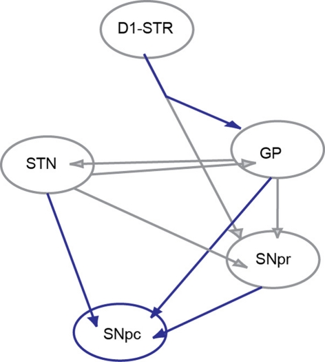
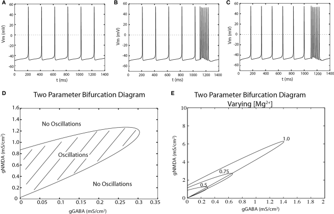
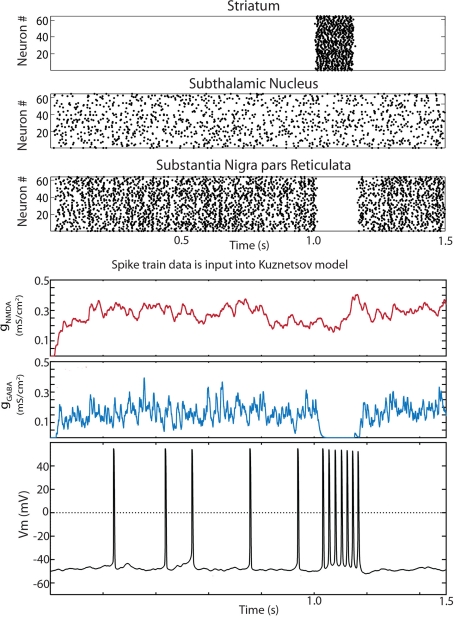
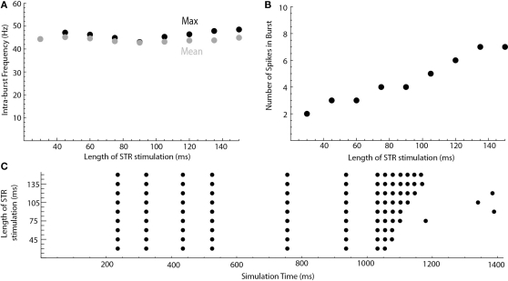
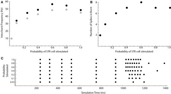
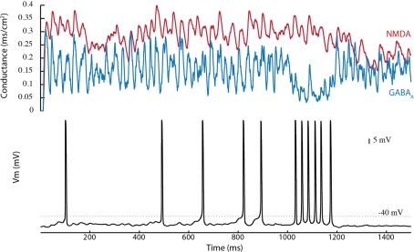
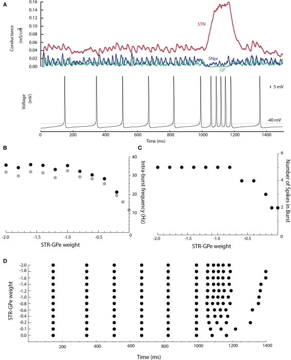
Substantia nigra pars compacta (SNpc) dopaminergic neurons receive strong tonic inputs from GABAergic neurons in the substantia nigra pars reticulata (SNpr) and globus pallidus (GP), and glutamatergic neurons in the subthalamic nucleus. The presence of these tonic inputs raises the possibility that phasic disinhibition may trigger phasic bursts in dopaminergic neurons. We first applied constant NMDA and GABA(A) conductances onto a two-compartment single cell model of the dopaminergic neuron (Kuznetsov et al., 2006). The model exhibited disinhibition bursting upon stepwise removal of inhibition. A further bifurcation analysis suggests that disinhibition may be more robust than excitation alone in that for most levels of NMDA conductance, the cell remains capable of bursting even after a complete removal of inhibition, whereas too much excitatory input will drive the cell into depolarization block. To investigate the network dynamics of disinhibition, we used a modified version of an integrate-and-fire based model of the basal ganglia (Humphries et al., 2006). Synaptic activity generated in the network was delivered to the two-compartment single cell dopaminergic neuron. Phasic activation of the D1-expressing medium spiny neurons in the striatum (D1STR) produced disinhibition bursts in dopaminergic neurons through the direct pathway (D1STR to SNpr to SNpc). Anatomical studies have shown that D1STR neurons have collaterals that terminate in GP. Adding these collaterals to the model, we found that striatal activation increased the intra-burst firing frequency of the disinhibition burst as the weight of this connection was increased. Our studies suggest that striatal activation is a robust means by which disinhibition bursts can be generated by SNpc dopaminergic neurons, and that recruitment of the indirect pathway via collaterals may enhance disinhibition bursting.

Keywords: GABA; burst; dopamine; model; network.


Figures

Figure 1
Figure 1
Circuit diagram. The six nuclei and their interconnections adapted from the Humphries et al. (2006) network model of the basal ganglia. The SNpc was added. The connections shown in gray are described in the original model while those in blue are added here. The connection from D1STR to the GP was added in accordance with anatomical data of Kawaguchi et al. (1990).
Figure 2
Figure 2
The dopaminergic neuron can fire disinhibition bursts. (A) Spontaneous, single-spiking in the absence of tonic input in a two-compartment model of the dopaminergic neuron. (B) Phasic activation of NMDA receptors (at t = 1100, 150 ms) produces a burst of action potentials. (C) The model exhibits single-spiking at similar frequencies as in (A) and can produces bursts by disinhibition upon complete removal of tonic inhibition at t = 1100 ms for 150 ms. (D) The NMDA and GABAA conductances at which repetitive spiking can be generated (hatched region) is computed by a two-point bifurcation diagram in AUTO (XPPAUT). (E) Magnesium dependence of the oscillatory region.
Figure 3
Figure 3
Disinhibition bursts can be evoked by striatal stimulation in the reduced Humphries et al. (2006) model. Raster plots for the 64 neurons in the D1 striatum, STN and SNpr. The only connections present in the reduced model were D1 striatum to SNpr, STN–SNpc, STN–SNpr, and SNpr–SNpc. At 1 s all 64 D1 striatal projection neurons were activated by current injection for a period of 150 ms. The simulation was run for a total of 10 s. The input to a random SNpc neuron was captured, saved to a file, and read in by the modified Kuznetsov et al. (2006) model. The synaptic conductances and resulting trace are shown below. Conductances were initially set at zero. The dopaminergic neuron model fired single spikes at approximately 5 Hz and exhibited a burst of action potentials during the period of striatal stimulation.
Figure 4
Figure 4
Length of striatal stimulation controls the number of spikes in the disinhibition burst but not the intra-burst firing frequency. (A) Shows the intra-burst firing frequency of the disinhibition burst (max. black, mean gray). (B) Shows the number of spikes in the burst. A raster plot of the results is shown in (C).
Figure 5
Figure 5
Weak striatal stimulation can cause disinhibition bursts. The probability that a striatal cell will be stimulated at t = 1000 ms in the reduced Humphries model is varied. (A) Shows the intra-burst firing frequency of the disinhibition burst (max. black, mean gray). (B) Shows the number of spikes in the burst. A raster plot of the results is shown in (C).
Figure 6
Figure 6
Tonic inhibition by GP reduces direct pathway mediated disinhibition bursting in the Humphries et al. (2006) model. In the Humphries model, tonic inhibition is provided by both the GP and SNpr. All connections are added except the D1STR–GP connection. Since GP and SNpr have similar firing rates, silencing of the SNpr by striatal stimulation removes approximately half of the inhibition at t = 1000 ms. Disinhibition bursts can still be produced.
Figure 7
Figure 7
The D1STR–GP connection promotes burst firing. (A) The D1STR–GP connection was added with a weight of −1.0. The synaptic conductances generated in the network model from the STN (red), GP (green), and SNpr (blue) are shown on the top. The resulting voltage trace is shown below. (B) Shows the intra-burst firing frequency of the disinhibition burst (max black, mean gray) as a function of the D1STR–GP weight. (C) Shows the number of spikes in the burst. A raster plot of the results is shown in (D). To avoid ceiling effects, in these simulations δ was reduced to δGABA = 0.0043 ms/cm2 and δNMDA = 0.0030 ms/cm2 so that the mean tonic conductances were approximately gGABAA = 0.02 ms/cm2 and gNMDA = 0.04 ms/cm2.

References

    1. Blythe S. N., Atherton J. F., Bevan M. D. (2007). Synaptic activation of dendritic AMPA and NMDA receptors generates transient high-frequency firing in substantia nigra dopamine neurons in vitro. J. Neurophysiol. 97, 2837–285010.1152/jn.01157.2006 - DOI - PubMed
    1. Blythe S. N., Wokosin D., Atherton J. F., Bevan M. D. (2009). Cellular mechanisms underlying burst firing in substantia nigra dopamine neurons. J. Neurosci. 29, 15531–1554110.1523/JNEUROSCI.2961-09.2009 - DOI - PMC - PubMed
    1. Brazhnik E., Shah F., Tepper J. M. (2008). GABAergic afferents activate both GABAA and GABAB receptors in mouse substantia nigra dopaminergic neurons in vivo. J. Neurosci. 28, 10386–1039810.1523/JNEUROSCI.2387-08.2008 - DOI - PMC - PubMed
    1. Charlety P. J., Grenhoff J., Chergui K., De la Chapelle B., Buda M., Svensson T. H., Chouvet G. (1991). Burst firing of mesencephalic dopamine neurons is inhibited by somatodendritic application of kynurenate. Acta Physiol. Scand. 142, 105–11210.1111/j.1748-1716.1991.tb09134.x - DOI - PubMed
    1. Chergui K., Akaoka H., Charléty P. J., Saunier C. F., Buda M., Chouvet G. (1994). Subthalamic nucleus modulates burst firing of nigra dopamine neurons via NMDA receptors. Neuroreport 5, 1185–1188 - PubMed