Review
. 2011 Sep;5(3):241-7.
doi: 10.1007/s12105-011-0269-8. Epub 2011 May 27.

Seromucinous hamartoma of the nasal cavity: a report of two cases and review of the literature

Affiliations
Review

Seromucinous hamartoma of the nasal cavity: a report of two cases and review of the literature

R A Khan et al. Head Neck Pathol. 2011 Sep.

Abstract

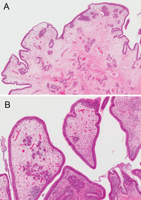
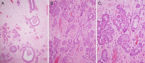
The sinonasal tract is a complex anatomic site, home to a wide variety of reactive, inflammatory, benign, and malignant lesions. Inflammatory polyps and papillomas are usually easily recognized by pathologists. A poorly understood lesion that has been more clearly defined in recent years is the nasal hamartoma. The epithelial subtypes include seromucinous hamartoma, respiratory epithelial adenomatoid hamartoma, and hybrid lesions. Seromucinous hamartomas have only been recognized and substantially reported over the past few years. They are a diagnostic challenge, needing to be distinguished from low grade adenocarcinomas, and are of interest because most of the basic questions about their pathophysiology remain unanswered. Herein, we present two novel cases of seromucinous hamartoma with features that partly expand the morphologic spectrum of these lesions, discuss the differential diagnosis, and review the literature to compare our findings with previously reported cases with the aim of better understanding this interesting entity.


Figures

Fig. 1
Fig. 1
Seromucinous hamartoma (case 1) showing a a complex, arborizing, polypoid lesion with a loose, pale stroma (20× magnification) with b a lining composed of ciliated respiratory epithelium overlying the submucosa containing scattered, bland, seromucinous glands (40× magnification)
Fig. 2
Fig. 2
Seromucinous hamartoma (case 1) showing a a loose submucosa with a mixture of bland seromucinous glands (left) and scattered invaginations of respiratory epithelium without pericellular basement membrane hyalinization (100× magnification). b Note the haphazard proliferation of seromucinous glands, which lack any epithelial tufting, back to back growth or cribriforming (200X magnification). c The seromucinous glands have scattered, eosinophilic zymogen granules and round, regular nuclei without atypia or mitotic activity (400× magnification)
Fig. 3
Fig. 3
Seromucinous hamartoma (Case 2) showing a a complex, polypoid lesion with more dense, eosinophilic submucosa (20× magnification) which has b abundant, haphazard seromucinous glands and scattered respiratory epithelial lined invaginations (40× magnification)
Fig. 4
Fig. 4
Seromucinous hamartoma (Case 2) showing a a fibrotic submucosa with a mixture of bland seromucinous glands and slightly cellular, fibrous stroma with bland spindle cells (100× magnification). b Focally, the seromucinous glands had a more atrophic lining with cells containing less cytoplasm but still retaining bland, round, hyperchromatic nuclei (200× magnification). c The seromucinous glands retained eosinophilic cytoplasmic zymogen granules and lacked any back to back growth, epithelial tufting, papillae or destructive growth (200× magnification)

References

    1. Ozolek JA, Hunt JL. Tumor suppressor gene alterations in respiratory epithelial adenomatoid hamartoma (REAH): comparison to sinonasal adenocarcinoma and inflamed sinonasal mucosa. Am J Surg Pathol. 2006;30(12):1576–1580. doi: 10.1097/01.pas.0000213344.55605.77. - DOI - PubMed
    1. Fusco A, Fedele M. Roles of HMGA proteins in cancer. Nat Rev Cancer. 2007;7(12):899–910. doi: 10.1038/nrc2271. - DOI - PubMed
    1. Perez-Ordonez B. Hamartomas, papillomas and adenocarcinomas of the sinonasal tract and nasopharynx. J Clin Pathol. 2009;62(12):1085–1095. doi: 10.1136/jcp.2007.053702. - DOI - PubMed
    1. Hsueh C, Hsueh S, Gonzalez-Crussi F, et al. Nasal chondromesenchymal hamartoma in children: report of 2 cases with review of the literature. Arch Pathol Lab Med. 2001;125(3):400–403. - PubMed
    1. McDermott MB, Ponder TB, Dehner LP. Nasal chondromesenchymal hamartoma: an upper respiratory tract analogue of the chest wall mesenchymal hamartoma. Am J Surg Pathol. 1998;22(4):425–433. doi: 10.1097/00000478-199804000-00006. - DOI - PubMed