. 2011 Oct 15;519(15):3085-101.
doi: 10.1002/cne.22667.

Genetic tracing of Nav1.8-expressing vagal afferents in the mouse

Affiliations

Genetic tracing of Nav1.8-expressing vagal afferents in the mouse

Laurent Gautron et al. J Comp Neurol. .

Abstract

Nav1.8 is a tetrodotoxin-resistant sodium channel present in large subsets of peripheral sensory neurons, including both spinal and vagal afferents. In spinal afferents, Nav1.8 plays a key role in signaling different types of pain. Little is known, however, about the exact identity and role of Nav1.8-expressing vagal neurons. Here we generated mice with restricted expression of tdTomato fluorescent protein in all Nav1.8-expressing afferent neurons. As a result, intense fluorescence was visible in the cell bodies, central relays, and sensory endings of these neurons, revealing the full extent of their innervation sites in thoracic and abdominal viscera. For instance, vagal and spinal Nav1.8-expressing endings were seen clearly within the gastrointestinal mucosa and myenteric plexus, respectively. In the gastrointestinal muscle wall, labeled endings included a small subset of vagal tension receptors but not any stretch receptors. We also examined the detailed innervation of key metabolic tissues such as liver and pancreas and evaluated the anatomical relationship of Nav1.8-expressing vagal afferents with select enteroendocrine cells (i.e., ghrelin, glucagon, GLP-1). Specifically, our data revealed the presence of Nav1.8-expressing vagal afferents in several metabolic tissues and varying degrees of proximity between Nav1.8-expressing mucosal afferents and enteroendocrine cells, including apparent neuroendocrine apposition. In summary, this study demonstrates the power and versatility of the Cre-LoxP technology to trace identified visceral afferents, and our data suggest a previously unrecognized role for Nav1.8-expressing vagal neurons in gastrointestinal functions.


Figures

Figure 1
Figure 1
A–I: Distribution of tdTomato fluorescence in sensory ganglia and central relays in the brainstem and spinal cord of Nav1.8-CretdTomato mice. Bright fluorescence was seen in whole mounts of the brainstem, including the NG (A,C) and its terminal field within the nucleus of the solitary tract (NTS; B,D). By using fluorescent microscopy, it is possible to recognize through the dorsal surface of the intact brainstem a heart-shaped fluorescent structure corresponding to the NTS (B). The NTS is also visible in a coronal slice shown in D. Similarly, it is possible to see fluorescence in whole mounts of the spinal cord, including the DRG (F) and its terminal field in the dorsal horn (G). Finally, the TG contained fluorescent neurons (H,I), whose central relay in the spinal trigeminal nucleus (sp5) is clearly visible in the intact brainstem (B,D,I). ap, Area postrema; dh, dorsal horn; JG, jugular ganglion; mand., mandibular branch of the V; max., maxillary branch of the V; ts, tract of the solitary nucleus; p, pituitary gland; PG, petrosal ganglion; opht., ophthalmic branch of the V; sp5, spinal trigeminal nucleus; TG, trigeminal ganglion; V, trigeminal nerve; X, vagus nerve. Scale bars ¼ 1 mm in A (applies to A,E,H); 500 μm in B (applies to B–D,F,G,I).
Figure 2
Figure 2
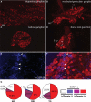
Characterization of tdTomato-positive neurons in sensory ganglia of Nav1.8-Cre mice. A large proportion of neurons was fluorescent in sections of the TG (A), vestibular/geniculate complex (B), NG (C), and DRG (D; epifluorescence and ApoTome). CGRP immunostaining (AlexaFluor-350) revealed that almost all CGRP neurons expressed tdTomato (E,F). Arrowheads in E and F indicate examples of CGRP neurons expressing tdTomato. Note that a few CGRP neurons in the DRG were tdTomato negative (arrows). The asterisks in F show examples of neuronal profiles being both CGRP- and tdTomato-negative. Pie charts showing the proportion of tdTomato- and CGRP-positive within examined sensory ganglia (G). CGRP-ir, calcitonin gene-related peptide immunoreactivity; DRG, dorsal root ganglion; GG, geniculate ganglion; NG, nodose ganglion; TG, trigeminal ganglion; VG, vestibular ganglion. Scale bar = 100 μm.
Figure 3
Figure 3
A–E: Additional description of tdTomato distribution in the vagus nerve and gut. Isolated neurons were seen in the SCG (whole mount; A) and the intestinal myenteric plexus (whole mount; D). The sensory component of the dorsovagal complex is heavily innervated by fibers originating from the nodose ganglion (cryostat-cut section; B). Fluorescent fibers can be discerned on a section cut through the cervical vagus nerve (C). Areas lacking fluorescence correspond to bundles of non-Cre-expressing fibers, including motor vagal neurons and certain mechanoreceptors. We observed the complete disappearance of vagal chemoreceptors in the mucosa after vagotomy (whole mount; D). ap, Area postrema; bv, blood vessel; DVC, dorsal vagal complex; nts, nucleus of the solitary tract; ts, tract of the solitary nucleus; SCG, superior cervical ganglion; VGX, vagotomy. Scale bars = 100 μm in A,B; 50 μm in C–E.
Figure 4
Figure 4
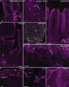
Examples of fluorescent fibers and terminals within selected tissues in Nav1.8-Cre-tdTomato mice. Bright native fluorescence (epifluorescence and ApoTome) was visible in various tissues innervated by sensory neurons, including the meninges (flattened whole mount; A), tongue (whole mount; B), muscle layers, submucosa and mucosa of the duodenum (C), airways and lungs (D), islets of Langherans (immunolabeled for glucagon-containing cells in green; E), inguinal lymph nodes (F), the skin (G), blood vessels of the epididymal fat (H), and muscle wall of the bladder (flattened whole mount; I). ad, Adipocyte; br, bronchioles; bv, blood vessel; d, dermis; ep, epidermis; fp, fongiform papilla; i, islet of Langerhans; mp, myenteric plexus; v, villi; WAT, white adipose tissue. Scale bars = 100 μm in A,C; 50 μm in B,G–I; 200 μm in D; 40 μm in E,F.
Figure 5
Figure 5
Reconstruction of the tongue innervation in Nav1.8-Cre-tdTomato mice. With the MosaiX module (Zeiss), we reconstructed the innervation of the tongue on a 16-μm cryostat-cut section (4×4 stitched tiles, five optical sections, 1-μm step). Bundles are seen reaching the epithelial surface to innervate individual taste buds. The inset shows a magnified view of a taste bud containing identified sensory endings. tb, Taste bud. Scale bar = 200 μm.
Figure 6
Figure 6
Detailed innervation of the mouse gallbladder and liver. Abundant innervation was seen in paravascular plexuses in the muscle wall of the gallbladder (A, whole mount; B, cryostat-cut section). Interestingly, vagotomy only partially reduced the innervation of the gall-blader, suggesting that spinal and vagal chemoreceptors are present in the gallbladder (C). Within hepatic lobes, we could clearly see varicose fibers endings in the vasculature of triads (D). It is also possible to see fluorescent fibers traveling in the portal vein wall (E) and in the adventia of bile ducts and hepatic artery at the hilar level (F). Arrows show representative fluorescent fibers. DAPI counterstaining is shown in D and F. bd, Bile duct; hA, hepatic artery; muc., mucosa; pv, portal vein; pvp, paravascular plexus; VGX, vagotomy. Scale bars = 50 μm in A; 40 μm in B; 20 μm in D; 100 μm in E,F.
Figure 7
Figure 7
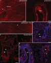
Identified specialized endings in Nav1.8-Cre-tdTomato mice. In whole mounts of the duodenum, tdTomato-positive fibers (epifluorescence and Apotome) are observed terminating the myenteric plexus forming specialized endings known as intraganglionic laminar endings (IGLEs; A,C). Arrows show representative IGLEs. One individual duodenal IGLE was reconstructed using Z optical sections (×40, 15 sections, 0.5-μm step) to show its structure better (c). A dense network of mucosal afferents is present in the villi along the entire intestinal tract (B). The typical innervation of an aortic body by chemoreceptors is represented in D. Note how a single vagal afferent enters the aortic body to form dense pericellular endings around glomus cells. In addition to IGLEs, thin fibers with varicosities circling around myenteric neurons were also seen in stomach and duodenum (C′,E,F). These fibers were generally positive for CGRP and were persistently seen after vagotomy (F), thus strongly suggesting their spinal origin. The example represented in E and F corresponds to the whole mount of the muscles layers of the stomach 4 days after bilateral subdiaphragmatic vagotomy. CGRP-ir, calcitonin gene-related peptide immunoreactivity; IGLEs, intraganglionic laminar endings; VGX, vagotomy. Scale bars = 100 μm in A,B,D; 25 μm in C (applies to C,C′); 50 μm in E (applies to E,F).
Figure 8
Figure 8
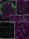
Anatomical proximity of Nav1.8-expressing mucosal afferents with select enteroendocrine cells. In the pancreas, tdTomato-positive fibers (epifluorescence and Apotome) are observed near glucagon-positive cells (AlexaFluor488; A). On occasion, varicose fibers were seen circling around glucagon cells, making apparent contacts (white arrows; A′,A′′). Typically, mucosal afferents in the duodenum form a dense network within the lamina propia (B) but rarely approach GLP-1-positive cells (AlexaFluor 488) very closely. In the stomach, ghrelin-positive cells (AlexaFluor 488) are distributed in crypts and lower parts of the mucosa (C). Ghrelin cells were frequently in the immediate vicinity of mucosal afferents (C,C′). Sometimes, apparent apposition between a ghrelin cell and an individual axon was observed (white arrow; C′). e, Epithelium; gm, gastric mucosa; GLP-1, glucagon-like peptide 1; i, islet of Langherans; lp, lamina propia. Scale bars = 20 μm.

References

    1. Abrahamsen B, Zhao J, Asante CO, Cendan CM, Marsh S, Martinez-Barbera JP, Nassar MA, Dickenson AH, Wood JN. The cell and molecular basis of mechanical, cold, and inflammatory pain. Science. 2008;321:702–705. - PubMed
    1. Akopian AN, Sivilotti L, Wood JN. A tetrodotoxin-resistant voltage-gated sodium channel expressed by sensory neurons. Nature. 1996;379:257–262. - PubMed
    1. Akopian AN, Souslova V, England S, Okuse K, Ogata N, Ure J, Smith A, Kerr BJ, McMahon SB, Boyce S, Hill R, Stanfa LC, Dickenson AH, Wood JN. The tetrodotoxin-resistant sodium channel SNS has a specialized function in pain pathways. Nat Neurosci. 1999;2:541–548. - PubMed
    1. Altschuler SM, Bao XM, Bieger D, Hopkins DA, Miselis RR. Viscerotopic representation of the upper alimentary tract in the rat: sensory ganglia and nuclei of the solitary and spinal trigeminal tracts. J Comp Neurol. 1989;283:248–268. - PubMed
    1. Berthoud HR, Neuhuber WL. Functional and chemical anatomy of the afferent vagal system. Auton Neurosci. 2000;85:1–17. - PubMed

Publication types

MeSH terms