. 2012 Aug;33(8):1868-82.
doi: 10.1002/hbm.21327. Epub 2011 May 26.

Brain activation for language dual-tasking: listening to two people speak at the same time and a change in network timing

Affiliations

Brain activation for language dual-tasking: listening to two people speak at the same time and a change in network timing

Augusto Buchweitz et al. Hum Brain Mapp. 2012 Aug.

Abstract

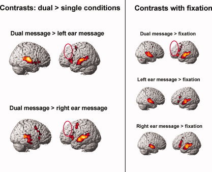
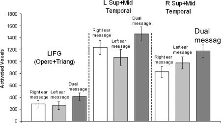
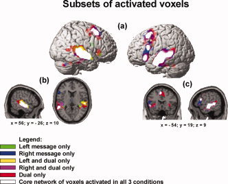
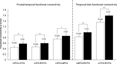
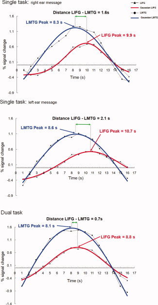
The study used fMRI to investigate brain activation in participants who were able to listen to and successfully comprehend two people speaking at the same time (dual-tasking). The study identified brain mechanisms associated with high-level, concurrent dual-tasking, as compared with comprehending a single message. Results showed an increase in the functional connectivity among areas of the language network in the dual task. The increase in synchronization of brain activation for dual-tasking was brought about primarily by a change in the timing of left inferior frontal gyrus (LIFG) activation relative to posterior temporal activation, bringing the LIFG activation into closer correspondence with temporal activation. The results show that the change in LIFG timing was greater in participants with lower working memory capacity, and that recruitment of additional activation in the dual-task occurred only in the areas adjacent to the language network that was activated in the single task. The shift in LIFG activation may be a brain marker of how the brain adapts to high-level dual-tasking.


Figures

Figure 1
Figure 1
Contrasts of dual task > single tasks and of each condition relative to fixation; clusters of voxels significant at P < 0.001, uncorrected, extent threshold = 6 voxels, T = 4.02. The dual task > right, dual task > left, and dual task > fixation contrasts show that there was no more activation in left DLPFC in dual than in the single tasks (red ellipses).
Figure 2
Figure 2
Activated voxels in single and dual tasks: more voxels activated in language‐network areas in dual‐tasking activated voxels for a threshold of T = 4.02 in the anatomically‐defined regions of the language network. LIFG is the mean of the activated voxels in the opercularis and triangularis parts of the inferior frontal gyrus [F3OP and F3T (Tzourio‐Mazoyer et al., 2002)]. The Temporal anatomical regions are the mean for the posterior and mid sections of T1 and T2, and HES (Tzourio‐Mazoyer et al., 2002).
Figure 3
Figure 3
Cortical areas activated in dual‐tasking: core network of shared voxels and the recruitment of areas circumscribing the core with increasing task difficulty clusters of voxels significant at P < 0.001, uncorrected, extent threshold = 6 voxels, T = 4.02: (a) surface rendering showing overlap of the three conditions with the core of voxels activated in all three conditions (intersection of voxels activated in Left Message, Right Message, and Dual Message); (b) sagittal view of the right‐hemisphere and axial view showing the subsets of activated voxels that circumscribe the core of commonly activated areas; (c) sagittal view of the lefthemisphere and coronal view showing the subsets of activated voxels. Dual task–only activation shows clusters that circumscribe the subset of voxels activated in the single tasks only and the intersection of voxels activated in the dual task and one of the single tasks (right and dual only or left and dual only).
Figure 4
Figure 4
Functional connectivity in dual‐tasking: higher synchronization between areas of the language network. Functional connectivity analyses for the language network regions (LIFG and bilateral mid and superior temporal). Increase in functional connectivity from single to dual condition was observed for the comparison between the FCA z‐score for the dual condition and the average FCA z‐score for the two single conditions (right and left ear message); (*) = P < 0.05; (**) = P < 0.01; (***) = P < 0.001.
Figure 5
Figure 5
Analysis of time courses: timing shift in lifg activation underpins synchronization of LIFG‐temporal brain activity time course plots for % signal change for the LIFG and LMTG2 functional regions of interest for one individual participant, averaged over individual trials. The peak coordinates (x‐axis) reported are for the Gaussian curve that represents the best fit calculated for the activation for this specific participant. The distance between the peak for the best‐fitting Gaussian curve for LIFG and LMTG decreases in the comparison between single and dual task.

References

    1. Chee MW, Tan EW, Thiel, T ( 1999): Mandarin and English single word processing studied with functional magnetic resonance imaging. J Neurosci 19: 3050–3056. - PMC - PubMed
    1. Chein JM, Schneider W ( 2005): Neuroimaging studies of practice‐related change: fMRI and meta‐analytic evidence of a domain‐general control network for learning. Brain Res Cogn Brain Res 25: 607–623. - PubMed
    1. Constable RT, Pugh KR, Berroya E, Mencl WE, Westerveld M, Ni W, Shankweiler D ( 2004): Sentence complexity and input modality effects in sentence comprehension: An fMRI study. Neuroimage 22: 11–22. - PubMed
    1. Costafreda SG, Fu CHY, Lee L, Everitt B, Brammer MJ, David AS ( 2006): A systematic review and quantitative appraisal of fMRI studies of verbal fluency: Role of the left inferior frontal gyrus. Hum Brain Mapp 27: 799–810. - PMC - PubMed
    1. Cox RW ( 1996): AFNI: Software for analysis and visualization of functional magnetic resonance neuroimages. Comput Biomed Res 29: 162–173. - PubMed

Publication types