

doi: 10.1021/nl201166k.
Epub 2011 May 27.
Controlled growth of nanoparticles from solution with in situ liquid transmission electron microscopy
Affiliations
- PMID: 21619024
- PMCID: PMC3162246
- DOI: 10.1021/nl201166k
Item in Clipboard
Controlled growth of nanoparticles from solution with in situ liquid transmission electron microscopy
Nano Lett.
.
Abstract
Direct visualization of lead sulfide nanoparticle growth is demonstrated by selectively decomposing a chemical precursor from a multicomponent solution using in situ liquid transmission electron microscopy. We demonstrate reproducible control over growth mechanisms that dictate the final morphology of nanostructures while observing growth in real-time with subnanometer spatial resolution. Furthermore, while an intense electron beam can initiate nanoparticle growth, it is also shown that a laser can trigger the reaction independently of the imaging electrons.
Figures

Bright Field and Dark Field Scanning TEM overviews of the parent PbS solution diluted 1,000x (a & d), 2,500x (b & e) and 5,000x (c & f) centered on an area previously scanned at higher magnification. All panels depict evidence of ion diffusion causing nucleation and growth of nanoparticles in a region surrounding the square of initial electron irradiation. Scale bars represent 500 nm (d & e) and 250 nm (f).
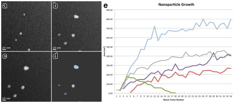
Correlated image series of the same area at time = 3.5 seconds (a), 15.1 seconds (b) and 26.8 seconds (c) showing the growth of individual PbS nanoparticles. (d) depicts the same image as (b) with individual nanoparticles identified with varying color. (e) plot of nanoparticle area versus time spanning 40 seconds with lines representing individual nanoparticles labeled according to color as in (d). Panels (a–c) correspond to frames 3, 13 and 23 respectively. Scale bars represent 20 nm.
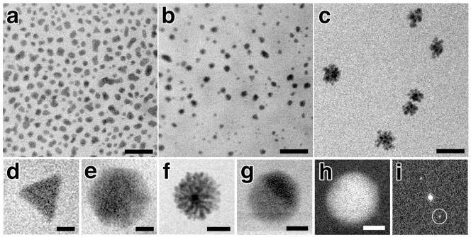
In situ Bright Field Scanning TEM images of 2:1 (a), 1:1 (b) and 1:1.25 (c) Pb:S solutions each at 5,000x dilution. Panels (d–g) show a gallery of differently shaped nanoparticles observed in situ including trigonal (d), hexagonal (e) flower-like (f), and spherical (g). Dark Field Scanning TEM image (h) of the same nanoparticle in (g) and the corresponding FFT (i). Lattice fringes for the (220) plane of PbS at 0.21 nm resolution can be seen in (g&h) and Bragg reflections circled in (i). Scale bars represent 100 nm (a–c), 12.5 nm (d&e), 25 nm (f), and 2.5 nm (g&h).

Correlated image series of the same area of a 1:1,000x dilution of 1:1 (Pb:S) solution illuminated solely with electron irradiation (a–c), or with electron irradiation but following a single sample drive laser pulse at an energy of 0.6 μj (d–f). For the electron irradiation only series, (a) corresponds to time = 0, while (b&c) are for time = 5 minutes and 15 minutes respectively. (d,e&f) correspond to the images 2 seconds, 2 minutes and 5 minutes after sample drive laser initiation, respectively. Arrow in (d) indicates first observed nuclei. Scale bars represent 3 μm.
References
Publication types
MeSH terms
Substances
Grants and funding
LinkOut - more resources
Full Text Sources
Other Literature Sources
