. 2011 May 27:9:32.
doi: 10.1186/1741-7007-9-32.

Real-time visualization of heterotrimeric G protein Gq activation in living cells

Affiliations

Real-time visualization of heterotrimeric G protein Gq activation in living cells

Merel J W Adjobo-Hermans et al. BMC Biol. .

Abstract

Background: Gq is a heterotrimeric G protein that plays an important role in numerous physiological processes. To delineate the molecular mechanisms and kinetics of signalling through this protein, its activation should be measurable in single living cells. Recently, fluorescence resonance energy transfer (FRET) sensors have been developed for this purpose.

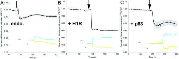
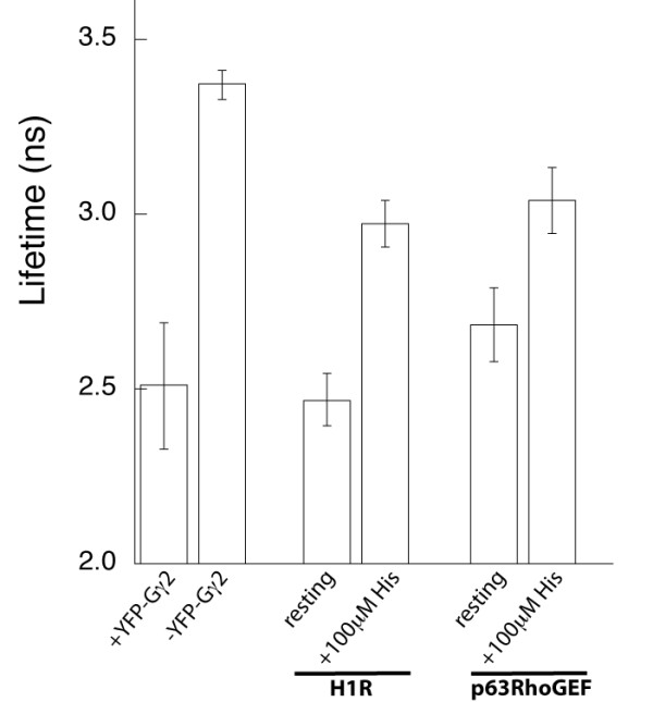
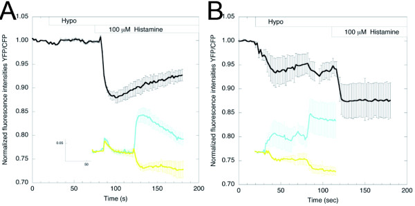
Results: In this paper, we describe the development of an improved FRET-based Gq activity sensor that consists of a yellow fluorescent protein (YFP)-tagged Gγ2 subunit and a Gαq subunit with an inserted monomeric Turquoise (mTurquoise), the best cyan fluorescent protein variant currently available. This sensor enabled us to determine, for the first time, the kon (2/s) of Gq activation. In addition, we found that the guanine nucleotide exchange factor p63RhoGEF has a profound effect on the number of Gq proteins that become active upon stimulation of endogenous histamine H1 receptors. The sensor was also used to measure ligand-independent activation of the histamine H1 receptor (H1R) upon addition of a hypotonic stimulus.

Conclusions: Our observations reveal that the application of a truncated mTurquoise as donor and a YFP-tagged Gγ2 as acceptor in FRET-based Gq activity sensors substantially improves their dynamic range. This optimization enables the real-time single cell quantification of Gq signalling dynamics, the influence of accessory proteins and allows future drug screening applications by virtue of its sensitivity.


Figures

Figure 1
Figure 1
Gαq-visible fluorescent protein (VFP) is functional. (A) HeLa cells expressing Gαq tagged with monomeric yellow fluorescent protein (Gαq-mYFP). (B) Mouse embryonic fibroblasts (MEFs) derived from wild-type mice showed an increase in [Ca2+]i upon addition (arrow) of bradykinin (BK) (1 μmol/l, n = 6. Error bars depict SE). (C) MEFs derived from Gαq/11-deficient mice (MEFq/11-/-) did not display increased [Ca2+]i upon addition of BK (n = 9). (D) Expression of wild-type Gαq in MEFq/11-/- caused an increase in cytosolic calcium upon addition of BK (n = 2). Most cells did not express the re-introduced wild-type (untagged) Gαq and did not show a decrease in Fura Red intensity upon addition of BK. (E) Expression of Gαq-mYFP in MEFq/11-/- caused an increase in cytosolic calcium upon addition of BK (n = 12); (Inset) MEFq/11-/- cell expressing Gαq-mYFP. (F) Gαq-mTqΔ6 similarly caused an increase in [Ca2+]i upon addition of BK (n = 6). (Inset) MEFq/11-/- cell expressing Gαq-mTqΔ6.
Figure 2
Figure 2
Dynamic range of Gq sensors. (A) The fluorescence resonance energy transfer (FRET) ratio change (yellow fluorescent protein:monomeric Turquoise (YFP:mTq)) upon addition of histamine (100 μmol/l) in HeLa cells coexpressing Gαq-mTqΔ6, Gβ1, YFP-Gγ2 and the histamine H1 receptor (H1R) (Inset) the mTq and YFP intensity traces from which the ratio was derived (n = 14; error bars depict SE). Addition of the H1R inverse agonist mepyramine (10 μmol/l) reversed the ratio change induced by histamine. (B) The FRET ratio change (YFP:CFP) upon addition of histamine (100 μmol/l) in HeLa cells coexpressing Gαq-enhanced (E)CFP, EYFP-Gβ1, Gγ2 and H1R. (Inset) the CFP and YFP intensity traces from which the YFP:CFP ratio was derived (n = 17; error bars depict SE). (C) Representative trace of the FRET ratio change (YFP:mTq) upon addition of histamine (100 μmol/l) in HeLa cells coexpressing Gαq- mTqΔ6, EYFP-Gβ1, Gγ2 and H1R. (Inset). The mTq and YFP intensity traces from which the ratio was derived. (D) The FRET ratio change (YFP:CFP) upon addition of histamine (100 μmol/l) in HeLa cells coexpressing Gαq-ECFP, Gβ1, YFP-Gγ2 and H1R. (Inset) the CFP and YFP intensity traces from which the ratio was derived (n = 5; error bars depict SE).
Figure 3
Figure 3
Dose-response curve. (A) Representative trace depicting the fluorescence resonance energy transfer (FRET) ratio changes (yellow fluorescent protein:monomeric Turquoise (YFP:mTq)) upon addition of increasing amounts of histamine (gray bars) in HeLa cells expressing Gαq-mTqΔ6, Gβ1, YFP-Gγ2 and the histamine H1 receptor (H1R). (B) The mTq and YFP intensity traces from which the ratio was derived.
Figure 4
Figure 4
Kinetics of Gq activation. (A) The fluorescence resonance energy transfer (FRET) ratio change (yellow fluorescent protein:monomeric Turquoise (YFP:mTq)) upon addition of histamine in cells expressing Gαq-mTqΔ6, Gβ1, YFP-Gγ2 and histamine H1 receptor (H1R), measured with high temporal resolution. Images were acquired at a frame rate of 0.064 seconds, and 1000 data points are shown. (B) Average FRET ratio change (scaled between 0 and 1) upon addition of histamine (n = 11; error bars depict SE). The data points were appropriately fitted with a monoexponential curve (solid line).
Figure 5
Figure 5
Accessory proteins affect the activation state of Gq. (A) endo.: the fluorescence resonance energy transfer (FRET) ratio change (yellow fluorescent protein:monomeric Turquoise (YFP:mTq)) upon addition (arrow) of histamine (100 μmol/l) in HeLa cells expressing Gαq-mTqΔ6, Gβ1 and YFP-Gγ2. (Inset) the mTq and YFP intensity traces that belong to the ratio (n = 9; error bars depict SE). (B) +H1R: the FRET ratio change (YFP/mTq) upon addition (arrow) of histamine (100 μmol/l) in HeLa cells expressing Gαq-mTqΔ6, Gβ1, YFP-Gγ2 and histamine H1 receptor (H1R). (Inset) the mTq and YFP intensity traces that belong to the ratio. (C) +p63: the FRET ratio change (YFP/mTq) upon addition of histamine (100 μmol/l) in HeLa cells expressing Gαq-mTqΔ6, Gβ1, YFP-Gγ2 and the guanine nucleotide exchange factor p63-RhoGEF. (Inset) the mTq and YFP intensity traces that belong to the ratio (n = 9).
Figure 6
Figure 6
Fluorescence lifetime data. Average cyan fluorescent protein (CFP) lifetimes measured under different conditions. The phase lifetime of Gαq-mTqΔ6 was measured in the absence (n = 15) and presence (n = 12) of the acceptor YFP-Gγ2. The difference in lifetime was significant. The Gq sensor was coexpressed with the histamine H1 receptor (H1R) and CFP lifetimes were measured before and after addition of 100 μmol/l histamine (n = 6). The Gq sensor was coexpressed with the guanine nucleotide exchange factor p63RhoGEF and CFP lifetimes were measured before and after addition of 100 μmol/l histamine (n = 6). In both conditions the lifetime difference before and after stimulation was significant. The CFP lifetimes before addition of histamine are not different (Student t-test, 95% CI) in the presence or absence of either H1R or p63RhoGEF.
Figure 7
Figure 7
Ligand-independent activation of Gq. (A) Graph depicting the fluorescence resonance energy transfer (FRET) ratio (yellow fluorescent protein:monomeric Turquoise (YFP:mTq)) change upon the application of a hypotonic (Hypo) stimulus, followed by the addition of histamine (100 μmol/l) in HeLa cells expressing Gαq-mTqΔ6, Gβ1 and YFP-Gγ2 (n = 5; error bars depict SE). (B) HeLa cells overexpressing the H1 receptor showed significant activation of Gq when the osmolality of the medium was reduced (Hypo). Subsequent addition of histamine yielded additional activation of Gq (n = 5).

References

    1. Cabrera-Vera TM, Vanhauwe J, Thomas TO, Medkova M, Preininger A, Mazzoni MR, Hamm HE. Insights into G protein structure, function, and regulation. Endocr Rev. 2003;24(6):765–781. doi: 10.1210/er.2000-0026. - DOI - PubMed
    1. Wall MA, Coleman DE, Lee E, Iniguez-Lluhi JA, Posner BA, Gilman AG, Sprang SR. The structure of the G protein heterotrimer Gi alpha 1 beta 1 gamma 2. Cell. 1995;83(6):1047–1058. doi: 10.1016/0092-8674(95)90220-1. - DOI - PubMed
    1. Taylor SJ, Chae HZ, Rhee SG, Exton JH. Activation of the beta 1 isozyme of phospholipase C by alpha subunits of the Gq class of G proteins. Nature. 1991;350(6318):516–518. doi: 10.1038/350516a0. - DOI - PubMed
    1. Booden MA, Siderovski DP, Der CJ. Leukemia-associated Rho guanine nucleotide exchange factor promotes G alpha q-coupled activation of RhoA. Mol Cell Biol. 2002;22(12):4053–4061. doi: 10.1128/MCB.22.12.4053-4061.2002. - DOI - PMC - PubMed
    1. Lutz S, Freichel-Blomquist A, Yang Y, Rumenapp U, Jakobs KH, Schmidt M, Wieland T. The guanine nucleotide exchange factor p63RhoGEF, a specific link between Gq/11-coupled receptor signaling and RhoA. J Biol Chem. 2005;280(12):11134–11139. doi: 10.1074/jbc.M411322200. - DOI - PubMed

Publication types

MeSH terms

Substances