. 2011 May 27;6(1):21.
doi: 10.1186/1749-8546-6-21.

The Yin and Yang actions of North American ginseng root in modulating the immune function of macrophages

Affiliations

The Yin and Yang actions of North American ginseng root in modulating the immune function of macrophages

Chike Godwin Azike et al. Chin Med. .

Abstract

Background: Immuno-modulatory effects of ginseng, including both immuno-stimulatory and immuno-suppressive effects, have been widely reported. This study aims to determine whether the paradoxical immuno-modulatory effect is related to unique phytochemical profiles of different North American (NA) ginseng, namely aqueous (AQ) and alcoholic (ALC) extracts.

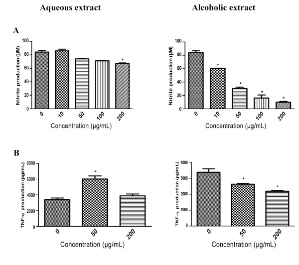
Methods: AQ and ALC extracts were prepared and their immuno-bioactivity were studied in vitro in murine macrophages (Raw 264.7) through measuring the direct stimulatory production of pro-inflammatory mediator and cytokines as well as the suppression of lipopolysaccharide (LPS)-stimulatory response by the two extracts. Gel permeation chromatography was used to fractionate and isolate phytochemicals for characterization of ginseng extracts.

Results: AQ extract up-regulated the production of nitric oxide (NO), tumour necrosis factor-α (TNF-α) and interleukin-6 (IL-6) while ALC extract did not. ALC extract but not AQ extract suppressed LPS-induced macrophage NO and TNF-α production. These immuno-stimulatory and suppressive effects were exhibited at similar extract concentrations. Moreover, the macrophage-stimulating activity of the AQ extract was inhibited in the presence of ALC extract. Fractionation of AQ extract revealed the presence of two major peaks at 230 nm with average molecular weights of 73,000 and 37,000 Da. The first fraction had similar elution volume as the crude polysaccharide (PS) fraction isolated from the AQ extract, and it was the only bioactive species. Parallel fractionation study of ALC extract yielded similar elution profiles; however, both sub-fractions were devoid of PS. Fraction I of the ALC extract suppressed LPS-induced NO production dose-dependently.

Conclusion: ALC extract of NA ginseng, which was devoid of PS, was immuno-inhibitory whereas the AQ extract, which contained PS, was immuno-stimulatory. These extract-related anti-inflammatory and pro-inflammatory effects may be considered as the Yin and Yang actions of ginseng.


Figures

Figure 1
Figure 1
Immuno-stimulatory effects of the AQ and ALC ginseng extracts on 24 hours macrophage production of (A) NO, (B) TNF-α and (C) IL-6. Murine macrophages (RAW 264.7 cells) were treated with or without AQ and ALC ginseng extracts (20, 50, 200 μg/ml), LPS (1 μg/ml) for 24 hours and the culture supernatants were analysed for NO and TNF-α/IL-6 by Griess reaction assay and ELISA respectively. Three independent experiments were performed and the data were shown as mean ± SD. Datasets were evaluated by ANOVA. * Values P < 0.001 compared to the untreated (vehicle) control were statistically significant.
Figure 2
Figure 2
Effect of the AQ and ALC ginseng extracts on LPS-stimulated 24 hours macrophage production of (A) NO and (B) TNF-α. Murine macrophages (RAW 264.7 cells) were pre-treated with without the AQ and ALC ginseng extracts (50, 200 μg/ml), for two hours after which LPS 1 μg/ml was added; and 24 hours later the NO and TNF-α contents of the culture supernatants were determined by Griess reaction assay and ELISA, respectively. Three independent experiments were performed and the data were shown as mean ± SD. Datasets were evaluated by ANOVA. * Values P < 0.001 compared to the LPS positive control were statistically significant.
Figure 3
Figure 3
ALC ginseng extract suppressed up-regulation of macrophage NO production by the AQ ginseng extract. Murine macrophages (RAW 264.7 cells) were pre-treated with or without the 200 μg/ml ALC ginseng extracts (50, 200 μg/ml), for two hours after which the AQ ginseng extract (50, 200 μg/ml) was added, and the NO contents of the culture supernatants were determined by Griess reaction assay 24 hours later. Three independent experiments were performed and the data were shown as mean ± SD. Datasets were evaluated by ANOVA. * Values P < 0.001 were statistically significant.
Figure 4
Figure 4
Sephadex G-75 (47 × 2.5 cm) chromatographic fractionation of the (A) AQ, (B) ALC and (C) PS extracts of ginseng. Column was loaded with 500 mg of extract, and then eluted with distilled water at flow rate of 1 mL/min. The y-axis is the absorbance at 230 nm while the x-axis represents the elution volume (mL).
Figure 5
Figure 5
Immuno-stimulatory effect of Fraction I and III of the AQ, PS extracts of ginseng and Cold-Fx. Murine macrophages (RAW 264.7 cells) were treated with Fraction I and III of AQ ginseng extract, PS extract of ginseng and Cold-Fx. (0, 20, 50, 200 μg/ml), LPS (1 μg/ml) for 24 hours and the NO, TNF-α and IL-6 contents of the culture supernatants were determined. Three independent experiments were performed and the data were shown as mean ± SD. Datasets were evaluated by ANOVA. *Values P < 0.001 compared to the untreated (vehicle) control were statistically significant.
Figure 6
Figure 6
Effect of Fractions I and III of the ALC extract on LPS-stimulated 24 hours macrophage production of NO. Murine macrophages (RAW 264.7 cells) were pre-treated with or without the AQ and ALC extracts (10, 50, 100, 200 μg/ml) for two hours after which LPS (1 μg/ml) was added, and the NO content of the culture supernatants were determined by Griess reaction assay 24 hours later. Three independent experiments were performed and the data were shown as mean ± SD. Datasets were evaluated by ANOVA. * Values P < 0.001 compared to the LPS positive control were statistically significant.
Figure 7
Figure 7
Identification of polysaccharides in Fraction I of the AQ extract, PS extract, AQ extract and ALC extract of ginseng by size exclusion chromatography. 100 μL of 5 mg/mL sample or 2.4 mg/mL standard was injected and eluted with 0.05 M NaNO3 mobile phase, this was monitored with right angle light scattering detector at 1 mL/min flow rate. Pullulan polysaccharide was used as a reference standard. The y-axis is the detector response (mV) while the x-axis represents the retention volume (mL). No signal was detected with the ALC extract, suggesting the absence of polysaccharides in this extract.

References

    1. Angelova N, Kong HW, van der Heijden R, Yang SY, Choi YH, Kim HK, Wang M, Hankemeier T, van der Greef J, Xu G, Verpoorte R. Recent methodology in phytochemical analysis of ginseng. Phytochem Anal. 2008;19:2–16. doi: 10.1002/pca.1049. - DOI - PubMed
    1. Borchers AT, Keen CL, Stern JS, Gershwin ME. Inflammation and native American medicine: the role of botanicals. Am J Clin Nutr. 2000;72:339–47. - PubMed
    1. Agriculture and Agri-Food Canada, Overview of the Canadian Special Crops Industry - Ginseng. http://www.ats-sea.agr.gc.ca/can/4752-eng.htm#j
    1. Health Canada Drugs and Health Products, Natural Health Product Monograph Panax Ginseng. http://www.hc-sc.gc.ca/dhp-mps/alt_formats/pacrb-dgapcr/pdf/prodnatur/ap...
    1. Attele AS, Wu JA, Yuan CS. Ginseng pharmacology; multiple constituents and multiple actions. Biochem Pharmacol. 1999;58:1685–1693. doi: 10.1016/S0006-2952(99)00212-9. - DOI - PubMed

LinkOut - more resources