. 2011 May 27:6:36.
doi: 10.1186/1750-1326-6-36.

Lack of a-disintegrin-and-metalloproteinase ADAM10 leads to intracellular accumulation and loss of shedding of the cellular prion protein in vivo

Affiliations

Lack of a-disintegrin-and-metalloproteinase ADAM10 leads to intracellular accumulation and loss of shedding of the cellular prion protein in vivo

Hermann C Altmeppen et al. Mol Neurodegener. .

Abstract

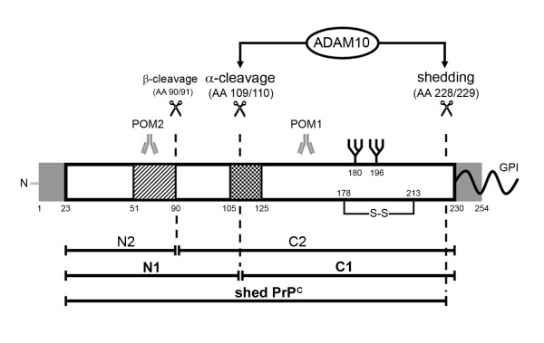
Background: The cellular prion protein (PrPC) fulfils several yet not completely understood physiological functions. Apart from these functions, it has the ability to misfold into a pathogenic scrapie form (PrPSc) leading to fatal transmissible spongiform encephalopathies. Proteolytic processing of PrPC generates N- and C-terminal fragments which play crucial roles both in the pathophysiology of prion diseases and in transducing physiological functions of PrPC. A-disintegrin-and-metalloproteinase 10 (ADAM10) has been proposed by cell culture experiments to be responsible for both shedding of PrPC and its α-cleavage. Here, we analyzed the role of ADAM10 in the proteolytic processing of PrPC in vivo.

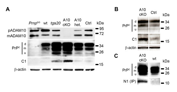
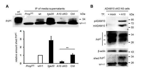
Results: Using neuron-specific Adam10 knockout mice, we show that ADAM10 is the sheddase of PrPC and that its absence in vivo leads to increased amounts and accumulation of PrPC in the early secretory pathway by affecting its posttranslational processing. Elevated PrPC levels do not induce apoptotic signalling via p53. Furthermore, we show that ADAM10 is not responsible for the α-cleavage of PrPC.

Conclusion: Our study elucidates the proteolytic processing of PrPC and proves a role of ADAM10 in shedding of PrPC in vivo. We suggest that ADAM10 is a mediator of PrPC homeostasis at the plasma membrane and, thus, might be a regulator of the multiple functions discussed for PrPC. Furthermore, identification of ADAM10 as the sheddase of PrPC opens the avenue to devising novel approaches for therapeutic interventions against prion diseases.


Figures

Figure 1
Figure 1
Proteolytic cleavage of PrPC. After cleavage of the N-terminal and GPI anchor signal peptides (grey), mature membrane-bound PrPC can undergo at least three different proteolytic cleavage events (scissors) in addition to phospholipase C cutting within its GPI anchor. While β-cleavage is mainly observed under pathological conditions, a role of ADAM10 is discussed in the context of both, constitutive α-cleavage and shedding of PrPC. The resulting fragments are shown underneath. Striped box: octameric repeat region; checked box: hydrophobic core domain. Epitopes of PrPC specific monoclonal antibodies POM1 and POM2 used in this study are indicated.
Figure 2
Figure 2
Increased amounts of PrPC but unaltered mRNA levels in ADAM10 cKO mice. (A) Western blot analysis for PrPC in primary neurons of Prnp0/0, wt, ADAM10 cKO and Nestin-Cre-negative littermate controls (all from E14 embryos). Quantification of PrPC amounts shows a statistically significant increase of PrPC in ADAM10 cKO neurons compared to littermate controls (p = 0,0013). All samples were normalized to wildtype which was set to one (n = 3 for wt and Prnp0/0; n = 5 for A10 cKO and Ctrl) (d, m, u = di-, mono-, unglycosylated PrPC; representative blot is shown). Immunohistochemical staining of PrPC in brains (B) and dorsal root ganglia (C) of E14 ADAM10 cKO mice and littermate controls shows enhanced immunosignal throughout the entire central nervous system with some degree of variation between individual neurons in ADAM10 cKO mice (C, scale bar: 20 μm). Brain of an age-matched Prnp0/0 mice was taken as negative control in B. (B, upper row: whole brain section, scale bar: 500 μm; bottom row: magnification of inserts, scale bar: 50 μm). (D) Quantitative RT-PCR for PrPC mRNA in brain homogenates of ADAM10 cKO and wildtype littermate controls shows no significant difference in expression levels between genotypes at E14 (n = 4) and P1 (n = 3).
Figure 3
Figure 3
PrPC accumulates in intracellular compartments in ADAM10 cKO neurons. (A) Confocal (upper two rows; scale bar overview: 100 μm, scale bar single neuron: 10 μm) and spinning disc (lower two rows; scale bar YZ-sections: 10 μm) immunofluorescence microscopy for PrPC in permeabilized wildtype neurons (Ctrl) shows PrPC in evenly distributed vesicular structures (open arrowhead). In ADAM10 cKO neurons, dense PrPC accumulations are detectable adjacent to the nucleus (filled arrowhead) and in neuronal processes. YZ-sections through individual neurons confirm these findings (arrow for PrPC accumulation in ADAM10 cKO). Asterisks mark nuclei. (B) Confocal microscopic analysis of co-stainings of PrPC (left column) with intracellular organelle marker proteins (middle column) in wildtype littermate (Control) and ADAM10 cKO neurons. Overlays are shown in the right column. Accumulations of PrPC are only found in ADAM10 cKO neurons (filled arrowheads in d, j, and p) and colocalize with protein disulfide isomerase (PDI) used as marker for ER (e, f) and Golgi marker GM130 (k, l). Partial colocalization of PrPC with the lysosomal marker LAMP1 is detectable in vesicular structures (q, r). Scale bars represent 10 μm. Asterisks in overlays mark nuclei.
Figure 4
Figure 4
No induction of p53 or p53-regulated pathways in ADAM10 cKO. (A) Western blot analysis in lysates of primary neurons from Prnp0/0, wt, tga20, ADAM10 cKO and littermate controls (all from E14 embryos) shows comparable protein amounts of p53. (B) Using quantitative RT-PCR, relative mRNA levels of p53 and its downstream signalling target Mdm2 were determined in brain homogenates of ADAM10 cKO mice and wildtype littermates controls at E14 (n = 4) and P1 (n = 3). No significant differences in expression levels of p53 and Mdm2 were observed between genotypes at both ages (E14: p = 0,75 for p53 and p = 0,44 for Mdm2; P1: p = 0,37 for p53 and p = 0,56 for Mdm2).
Figure 5
Figure 5
ADAM10 is not responsible for α-cleavage of PrPC. (A) Western blot analysis for premature (pADAM10) and mature ADAM10 (mADAM10; upper row), PrPC (second row), PrPC C1 fragment after PNGase F treatment (third row), and actin (bottom row) in neuronal cultures from Prnp0/0, wt, tga20, ADAM10 cKO and littermate controls at E14. Levels of ADAM10 are dramatically reduced in ADAM10 cKO when compared to cultures from littermate controls or from mice overexpressing or lacking PrPC. In accordance with figure 2, PrPC levels are increased in ADAM10 cKO and there is no influence of ADAM10 status on the presence of the C1 fragment, which is most abundant in neuronal lysates with elevated expression of PrPC (i.e. ADAM10 cKO and tga20). (B) Western blot analysis for PrPC in brain of ADAM10 cKO and littermate controls at E14 confirms increased PrPC and the presence of the C1 fragment in ADAM10 cKO mice in an in vivo setting. (C) Western blot analysis for PrPC in lysates of ADAM10 cKO and wildtype neurons (both from E14 mice embryos) confirms increased PrPC amounts in the absence of ADAM10. Soluble N1 fragment was immunoprecipitated (IP) from culture supernatants of these neurons and is found to be increased in supernatants derived from ADAM10 cKO neurons.
Figure 6
Figure 6
ADAM10 is responsible for the shedding of PrPC. (A) Shed PrPC was immunoprecipitated (IP) from culture supernatants of primary neurons derived from Prnp0/0, wt, tga20, ADAM10 cKO and littermate control mice (all from E14 embryos) and visualized by Western blot analysis for PrPC. Shed PrPC, which shows a 2-3 kDa shift when compared to PrPC in lysates, is only detectable in wt, tga20, and littermate control neurons. Supernatants from ADAM10 cKO neuron cultures contain virtually no shed PrPC. IgG light chain (IgG-LC) of capture antibody is detectable at 25 kDa (representative blot is shown). Quantification confirms a significant reduction (p = 0,0026) of the amounts of shed PrPC shedding in ADAM10 cKO compared to littermate control cultures (Prnp0/0 values were defined as background and subtracted from the values of all other genotypes; wildtype values were set to one; n = 3 for tga20; n = 5 for A10 cKO and Ctrl). (B) Neural stem (NS) cells derived from an ADAM10 cKO mouse embryo at E14 were transfected (TF) without (+mock) or with Adam10-ORF (+A10). Following neuronal differentiation, PrPC was immunoprecipitated from culture supernatants (bottom row; IgG-LC = IgG light chain of capture antibody) and detected in corresponding cell lysates (second row; d, m, u = di-, mono-, unglycosylated PrPC) by Western blotting. Shed PrPC reappears in Adam10- but not in mock-transfected cultures (bottom row). Success of transfection was verified by Western blot detection of pADAM10 and mADAM10 in cell lysates (top row). Actin is shown as control (third row).

References

    1. Steele AD, Emsley JG, Ozdinler PH, Lindquist S, Macklis JD. Prion protein (PrPc) positively regulates neural precursor proliferation during developmental and adult mammalian neurogenesis. Proc Natl Acad Sci USA. 2006;103(9):3416–3421. doi: 10.1073/pnas.0511290103. - DOI - PMC - PubMed
    1. Bremer J, Baumann F, Tiberi C, Wessig C, Fischer H, Schwarz P, Steele AD, Toyka KV, Nave KA, Weis J. et al.Axonal prion protein is required for peripheral myelin maintenance. Nat Neurosci. 2010;13(3):310–318. doi: 10.1038/nn.2483. - DOI - PubMed
    1. Sunyach C, Cisse MA, da Costa CA, Vincent B, Checler F. The C-terminal products of cellular prion protein processing, C1 and C2, exert distinct influence on p53-dependent staurosporine-induced caspase-3 activation. J Biol Chem. 2007;282(3):1956–1963. - PubMed
    1. Chesebro B, Race B, Meade-White K, Lacasse R, Race R, Klingeborn M, Striebel J, Dorward D, McGovern G, Jeffrey M. Fatal transmissible amyloid encephalopathy: a new type of prion disease associated with lack of prion protein membrane anchoring. PLoS Pathog. 2010;6(3):e1000800. doi: 10.1371/journal.ppat.1000800. - DOI - PMC - PubMed
    1. Klingeborn M, Race B, Meade-White KD, Rosenke R, Striebel JF, Chesebro B. Crucial Role for Prion Protein Membrane Anchoring in the Neuroinvasion and Neural Spread of Prion Infection. J Virol. 2010. - PMC - PubMed