. 2011 May 28:12:119.
doi: 10.1186/1471-2474-12-119.

A prospective study of shoulder pain in primary care: prevalence of imaged pathology and response to guided diagnostic blocks

Affiliations

A prospective study of shoulder pain in primary care: prevalence of imaged pathology and response to guided diagnostic blocks

Angela Cadogan et al. BMC Musculoskelet Disord. .

Abstract

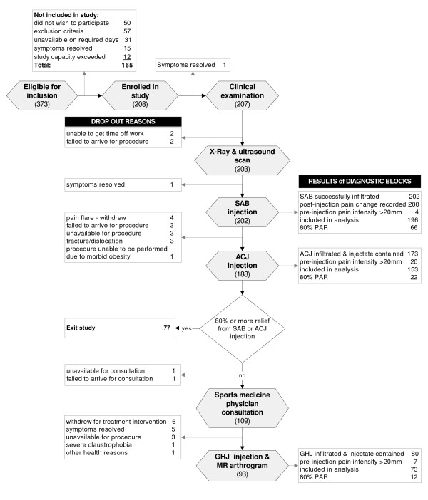
Background: The prevalence of imaged pathology in primary care has received little attention and the relevance of identified pathology to symptoms remains unclear. This paper reports the prevalence of imaged pathology and the association between pathology and response to diagnostic blocks into the subacromial bursa (SAB), acromioclavicular joint (ACJ) and glenohumeral joint (GHJ).

Methods: Consecutive patients with shoulder pain recruited from primary care underwent standardised x-ray, diagnostic ultrasound scan and diagnostic injections of local anaesthetic into the SAB and ACJ. Subjects who reported less than 80% reduction in pain following either of these injections were referred for a magnetic resonance arthrogram (MRA) and GHJ diagnostic block. Differences in proportions of positive and negative imaging findings in the anaesthetic response groups were assessed using Fishers test and odds ratios were calculated a for positive anaesthetic response (PAR) to diagnostic blocks.

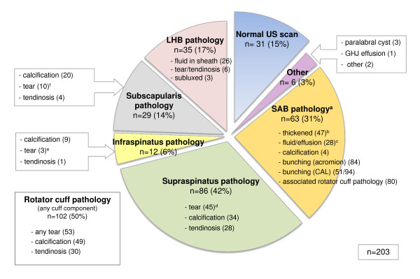
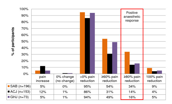
Results: In the 208 subjects recruited, the rotator cuff and SAB displayed the highest prevalence of pathology on both ultrasound (50% and 31% respectively) and MRA (65% and 76% respectively). The prevalence of PAR following SAB injection was 34% and ACJ injection 14%. Of the 59% reporting a negative anaesthetic response (NAR) for both of these injections, 16% demonstrated a PAR to GHJ injection. A full thickness tear of supraspinatus on ultrasound was associated with PAR to SAB injection (OR 5.02; p < 0.05). Ultrasound evidence of a biceps tendon sheath effusion (OR 8.0; p < 0.01) and an intact rotator cuff (OR 1.3; p < 0.05) were associated with PAR to GHJ injection. No imaging findings were strongly associated with PAR to ACJ injection (p ≤ 0.05).

Conclusions: Rotator cuff and SAB pathology were the most common findings on ultrasound and MRA. Evidence of a full thickness supraspinatus tear was associated with symptoms arising from the subacromial region, and a biceps tendon sheath effusion and an intact rotator cuff were associated with an intra-articular GHJ pain source. When combined with clinical information, these results may help guide diagnostic decision making in primary care.


Figures

Figure 1
Figure 1
Distribution of primary pain required for inclusion in the study.
Figure 2
Figure 2
Diagram showing study procedures, results of diagnostic blocks and dropout explanations. SAB, subacromial bursa; PAR, positive anaesthetic response (≥80% post-injection reduction in pain intensity); ACJ, acromioclavicular joint; GHJ, glenohumeral joint; MR arthrogram, magnetic resonance arthrogram. Numbers refer to the number (n) of subjects.
Figure 3
Figure 3
Prevalence of pathology identified on x-ray. n, number of cases; ACJ, acromioclavicular joint; GHJ, glenohumeral joint
Figure 4
Figure 4
Prevalence of pathology identified on ultrasound scan. (n), number of cases; US, ultrasound; GHJ, glenohumeral joint; SAB, subacromial bursa; CAL, coracoacromial ligament; LHB, long head of biceps tendon. aSubacromial pathology: any one of three present; dimension ≥2 mm, fluid/effusion or calcification. bSubacromial bursa dimensions: <1 mm (71); 1-2 mm (82); 2-3 mm (42); >3 mm (5). cSubacromial bursal effusion associated with full thickness rotator cuff tear (7). dSupraspinatus tears: intrasubstance (23); partial thickness-bursal surface (4); partial thickness-articular surface (8); full thickness (10). eInfraspinatus tears: intrasubstance (1); partial thickness (1); full thickness (1). fSubscapularis tears: intrasubstance (5); partial thickness (4); full thickness (1).
Figure 5
Figure 5
Shoulder x-ray images of ACJ pathology and rotator cuff calcification. a) AP x-ray view in external rotation showing degenerative acromioclavicular joint changes (white arrow); b) outlet view showing calcification in line with the infraspinatus tendon (black arrow).
Figure 6
Figure 6
Ultrasound scan images of subacromial bursa and supraspinatus pathology. a) hypoechoic region (between calipers) indicating an intrasubstance tear within posterior fibres of supraspinatus (longitudinal view) overlying the head of humerus (white arrowhead); b) thickened subacromial bursa (calipers); c) bunching of the SAB (white arrow) under the acromion during dynamic abduction.
Figure 7
Figure 7
Prevalence of subacromial bursa bunching under the acromion and coracoacromial ligament on ultrasound during dynamic abduction. SAB, subacromial bursa; US, ultrasound; CAL, coracoacromial ligament. Percentages are in reference to the number of cases in which bursal bunching was assessed (acromion n = 195; CAL n = 94).
Figure 8
Figure 8
Prevalence of pathology identified on MR arthrogram. (n), number of cases; LHB, long head of biceps tendon; ACJ, acromioclavicular joint; GHJ, glenohumeral joint; OA, osteoarthritis; SAB, subacromial bursa; aACJ degenerative changes: mild (28); moderate (18); severe (5). bAcromioclavicular joint pathology - other: os acromiale (2); unfused acromial ossification centre (1); acromial spur (4); widened joint space/subluxation (2); synovitis (1). cRotator interval pathology: coracohumeral or superior glenohumeral ligament thickening (40); rotator interval synovitis (39); biceps pulley, coracohumeral or superior glenohumeral ligament tear (13). dGlenoid labrum tear: isolated labral tear (5); associated pathology present (39); SLAP tear (20); SLAP Type II (17), Type III (2), Type IV (1); anterior-inferior tear (9); semi- or full circumferential tear (7); posterior-superior tear (1); other tear (9); paralabral cyst (10); paralabral cyst causing suprascapular nerve compression (2). eGlenohumeral joint pathology - other: bony irregularity humeral head without marrow oedema (12); Hill-Sachs lesion (3); intra-articular/osseous body (3); ganglion cyst between coracoacromial and coracohumeral ligaments (1); greater tuberosity fracture (1). fSubacromial bursitis: mild (52); moderate (12); severe (4) gSupraspinatus tears: intrasubstance (11); partial thickness-bursal surface (5); partial thickness articular surface (12); full thickness (7). hInfraspinatus tears: intrasubstance (4); partial thickness (3); full thickness (0) iSubscapularis tears: intrasubstance (4); partial thickness (0); full thickness (2)
Figure 9
Figure 9
MR arthrogram images of shoulder pathology. a) subacromial bursitis - coronal PD fat saturated image showing region of hyperintensity in the subacromial bursa (black arrow); b) partial thickness, articular surface supraspinatus tear (white arrow) - coronal T1 fat saturated image showing contrast extending into the supraspinatus tendon. c) ACJ degenerative changes (white arrow) -coronal PD fat saturated image; d) type III SLAP tear (white arrow) with contrast filling a paralabral cyst (black arrow) which extended into the supraglenoid and suprascapular notch causing neural compression -coronal PD fat saturated image.
Figure 10
Figure 10
Anaesthetic responses to diagnostic blocks. SAB, subacromial bursa; ACJ, acromioclavicular joint; GHJ, glenohumeral joint.

Similar articles

Cited by

References

    1. van der Windt DAWM, Koes BW, De Jong BA, Bouter LM. Shoulder disorders in general practice: Incidence, patient characteristics, and management. Ann Rheum Dis. 1995;54(12):959–964. doi: 10.1136/ard.54.12.959. - DOI - PMC - PubMed
    1. Luime JJ, Koes BW, Hendriksen IJM, Burdorf A, Verhagen AP, Miedema HS, Verhaar JA. Prevalence and incidence of shoulder pain in the general population: a systematic review. Scand J Rheumatol. 2004;33(2):73–81. doi: 10.1080/03009740310004667. - DOI - PubMed
    1. Croft PR, Pope DP, Silman AJ. The clinical course of shoulder pain: prospective cohort study in primary care. Br Med J. 1996;313(7057):601–602. - PMC - PubMed
    1. Urwin M, Symmons D, Allison T, Brammah T, Busby H, Roxby M, Simmons A, Williams G. Estimating the burden of musculoskeletal disorders in the community: the comparative prevalence of symptoms at different anatomical sites, and the relation to social deprivation. Ann Rheum Dis. 1998;57:649–655. doi: 10.1136/ard.57.11.649. - DOI - PMC - PubMed
    1. van der Windt DAWM, Koes BW, Bocke AJP, Deville W, de Jong BA, Bounter LM. Shoulder disorders in general practice: prognostic indicators of outcome. Br J Gen Pract. 1996;46:519–523. - PMC - PubMed

Publication types

MeSH terms