. 2011 Aug 15;317(14):1955-69.
doi: 10.1016/j.yexcr.2011.05.009. Epub 2011 May 18.

The STAT3 beacon: IL-6 recurrently activates STAT 3 from endosomal structures

Affiliations

The STAT3 beacon: IL-6 recurrently activates STAT 3 from endosomal structures

Christopher L German et al. Exp Cell Res. .

Abstract

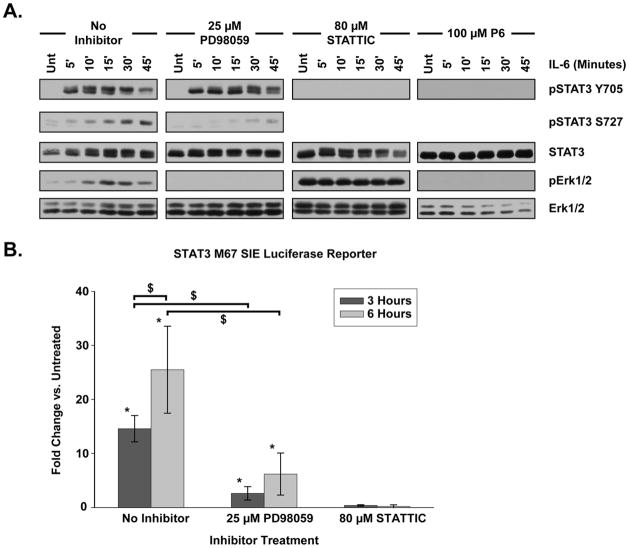
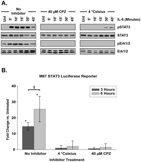
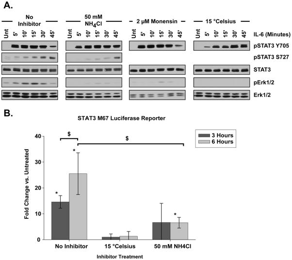
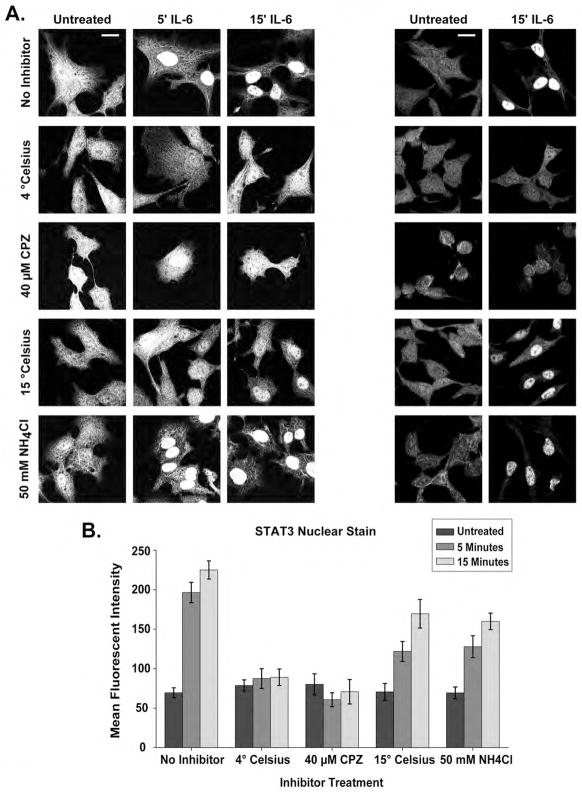
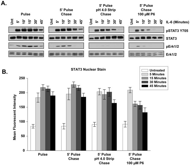
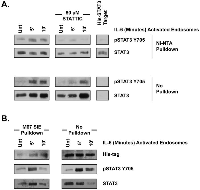
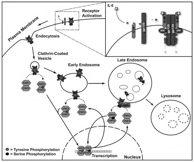
Endocytic trafficking plays an important role in signal transduction. Signal transducer and activator of transcription 3 (STAT3) and mitogen-activate protein kinase (MAPK) have both been localized to endosomal structures and are dependent upon endocytosis for downstream function. While the dependence of MAPK signaling upon endosomes has been well characterized, the involvement of endosomes in regulating STAT3 signaling has not been defined. Consequently, this study evaluated the role of endosomes in the initiation, modulation, amplification and persistence of interleukin-6(IL-6)-induced STAT3 signal transduction and transcription, and utilized IL-6-induced MAPK signaling as a comparator. Using pharmacologic treatment and temperature control of endocytic trafficking, pulse-chase treatments and in vitro kinase assays, STAT3 was found to interact with endosomes in a markedly different fashion than MAPK. STAT3 was activated by direct interaction with internal structures upstream of the late endosome following IL-6 exposure and persistent STAT3 signaling depended upon recurrent activation from endocytic structures. Further, STAT3 subcellular localization was not dependent upon endocytic trafficking. Instead, STAT3 transiently interacted with endosomes and relocated to the nucleus by an endosome-independent mechanism. Finally, endocytic trafficking played a central role in regulating STAT3 serine 727 phosphorylation through crosstalk with the MAPK signaling system. Together, these data reveal endosomes as central to the genesis, course and outcome of STAT3 signal transduction and transcription.


Figures

Figure 1
Figure 1. IL-6 Activates STAT3 and Erk1/2
(A) HepG2 cells were treated with IL-6 (20 ng/mL) for the indicated period of time. STAT3 and Erk1/2 activation was assessed by phosphorylation profile. The MEK1/2 inhibitor PD 98059, the STAT3 inhibitor STATTIC, and the broad spectrum JAK inhibitor P6 were all applied for 15 minutes prior to IL-6 treatment. Blots shown are representative of at least three separate, replicate experiments. (B) M67 SIE luciferase reporter activity in HepG2 cells treated with IL-6 for 3 or 6 hours. Cells were incubated with inhibitors for 15 minutes prior to IL-6 treatment. Fold change is compared against untreated samples. PD98059 treated cells exhibited a significant reduction in luciferase activity as compared to cells without inhibitor at both 3 and 6 hours post-IL-6. No significant luciferase activity was detected in STATTIC treated cells at any time period. Data were analyzed by two-way ANOVA with Student-Neuman-Keuls (SNK) pairwise comparison. Error bars indicate 95% confidence intervals. * indicates significant difference from untreated, p < 0.05. $ indicates significant difference between specified treatments, p < 0.05.
Figure 2
Figure 2. STAT3 Associates with Vesicular Structures
Blots shown are characteristic of results seen within at least three separate and replicate experiments. (A) Subcellular fractions isolated from IL-6 (20 ng/mL) treated HepG2 cells using Balch homogenization and differential centrifugation as described within the materials and methods section. (B) Transmission electron microscopy of the combined 16,000 x g and 115,000 x g “endosomes” fraction after phosphotungstic acid negative staining, showing and enriched population of membrane-bound organelles. (C) HepG2 cells were treated with IL-6 (20 ng/mL) for the indicated period of time and subcellular fractions were isolated. The endosomes and nuclear fractions were prepared as described and incubated with a biotinylated M67 SIE oligonucleotide to test the DNA binding capacity of STAT3 within these fractions. Protein bound to DNA was separated by pull down with neutravidin beads and identified as ‘SIE Pulldown’. Protein that did not bind DNA and precipitate with neutravidin beads is identified as ‘No Pulldown’.
Figure 3
Figure 3. STAT3 Requires Endocytosis for Activation
(A) Phosphorylation profile of STAT3 and Erk1/2 following IL-6 treatment (20 ng/mL) for the indicated period of time. Cells were treated with clathrin-mediated internalization inhibitor chlorpromazine (CPZ) or held at 4° C for 15 minutes prior to IL-6 application. Three separate replicate experiments are represented by the blots shown. (B) M67 SIE luciferase reporter activity in IL-6 treated cells that were held at 4° C, inhibited with CPZ, or treated without inhibitors for the period of time indicated. Inhibition and temperature manipulation was applied for 15 minutes prior to IL-6 treatment. Fold change is compared against cells not treated with IL-6. Neither 4 °C restricted or CPZ treated cells exhibited a significant increase in luciferase activity as compared to untreated cells either 3 or 6 hours following IL-6 treatment. Data were analyzed by two-way ANOVA and pairwise comparisons were made using the SNK method. Error bars indicate 95% confidence intervals. * indicates significantly different from untreated, p < 0.05. $ indicates significant difference between specified treatments, p < 0.05.
Figure 4
Figure 4. STAT3 is Activated from Endocytic Structures
(A) STAT3 and Erk1/2 phosphorylation profile after IL-6 treatment (20 ng/mL) for the indicated time period. Disruption of endocytic trafficking was achieved by altering vesicular pH with ammonium chloride (NH4Cl) or monensin treatment, or holding cells at 15° C for 15 minutes prior to IL-6 application. Blots shown are representative of at least three separate replicate experiments. (B) M67 SIE luciferase reporter activity in cells treated with IL-6 for the amount of time indicated. Inhibition or temperature manipulation was done for 15 minutes prior to IL-6 application. Fold change is compared to cells untreated with IL-6. No significant increase in luciferase activity over untreated controls was detected in 15 °C restricted cells at either 3 or 6 hours following IL-6 treatment or in NH4Cl treated cells 3 hours after IL-6 treatment. NH4Cl treated cells exhibited a significant increase in luciferase activity over untreated cells 6 hours post-IL-6, but this level of activity was significantly when compared to cells without inhibitor. Error bars indicate 95% confidence intervals and data were analyzed by two-way ANOVA with pairwise comparisons made using the SNK method. * indicates significantly different from untreated, p < 0.05. $ indicates significant difference between specified treatments, p < 0.05.
Figure 5
Figure 5. STAT3 Rapidly Localizes to the Nucleus Following IL-6 Treatment
(A) STAT3 staining within HepG2 cells under various inhibitor treatments and temperature restriction following IL-6 treatment. Panels on the left demonstrate STAT3 staining in both the nuclear and cytoplasmic compartments. Panels on the right demonstrate fields from which mean fluorescent intensity of STAT3 nuclear staining was calculated. (B) Mean fluorescent intensity of STAT3 nuclear staining under the treatment conditions in (A) calculated as described in the Experimental Procedues. Error bars represent 95% confidence intervals and significance is discussed within the Results section. Scale bars are equivalent to 20 um.
Figure 6
Figure 6. Persistent STAT3 Activation Arises from Endocytic Structures
(A) STAT3 and Erk1/2 phosphorylation profile following pulse or pulse-chase 20 ng/mL IL-6 treatment as described in the Experimental Procedures and the Results sections. Blots shown are representative of results seen in at least three independent experiments. (B) Mean fluorescent intensity of STAT3 nuclear staining across a time course of 20 ng/mL IL-6 pulse and pulse-chase conditions. Error bars indicate 95% confidence intervals and significance is discussed within the Results section.
Figure 7
Figure 7. Endosomes Directly Activate STAT3
(A) Phosphorylation profile of His-STAT3 by in vitro kinase assay. Endosomes isolated from HepG2 cells treated with 20 ng/mL IL-6 as indicated were mixed with His-tagged recombinant human STAT3 target for 30 minutes at 30 C. Ni+-NTA pulldown indicates protein isolated with Ni+-NTA agarose beads. No pulldown indicates protein that did not bind Ni+-NTA beads. Blots shown are representeative of at least three, replicate experiments. (B) M67 SIE oligo binding following an endosome in vitro kinase assay as in (A). At the conclusion of the kinase assay, 500 ng biotinylated M67 SIE oligo was used to pull down protein with DNA binding capacity as described within experimental procedures. ‘Pulldown’ indicates protein bound to the biotinylated oligo and isolated with neutravidin beads. ‘No Pulldown’ indicates protein that did not bind the biotinylated oligo.
Figure 8
Figure 8
Model of Endocytic Regulation of IL-6-Induced STAT3 Signaling.

References

    1. Howe CL, Mobley WC. Long-distance retrograde neurotrophic signaling. Curr Opin Neurobiol. 2005;15:40–48. - PubMed
    1. Howe CL. Modeling the signaling endosome hypothesis: why a drive to the nucleus is better than a (random) walk. Theor Biol Med Model. 2005;2:43. - PMC - PubMed
    1. Delcroix JD, Valletta JS, Wu C, Hunt SJ, Kowal AS, Mobley WC. NGF signaling in sensory neurons: evidence that early endosomes carry NGF retrograde signals. Neuron. 2003;39:69–84. - PubMed
    1. Beattie EC, Zhou J, Grimes ML, Bunnett NW, Howe CL, Mobley WC. A signaling endosome hypothesis to explain NGF actions: potential implications for neurodegeneration. Cold Spring Harb Symp Quant Biol. 1996;61:389–406. - PubMed
    1. Howe CL, Mobley WC. Signaling endosome hypothesis: A cellular mechanism for long distance communication. J Neurobiol. 2004;58:207–216. - PubMed

Publication types

MeSH terms

Substances