

Acute joint pathology and synovial inflammation is associated with increased intra-articular fracture severity in the mouse knee
- PMID: 21619936
- PMCID: PMC3312469
- DOI: 10.1016/j.joca.2011.04.011
Acute joint pathology and synovial inflammation is associated with increased intra-articular fracture severity in the mouse knee
Abstract
Objective: Post-traumatic arthritis is a frequent cause of disability and occurs most commonly and predictably after articular fracture. The objective of this investigation was to examine the effect of fracture severity on acute joint pathology in a novel murine model of intra-articular fracture.
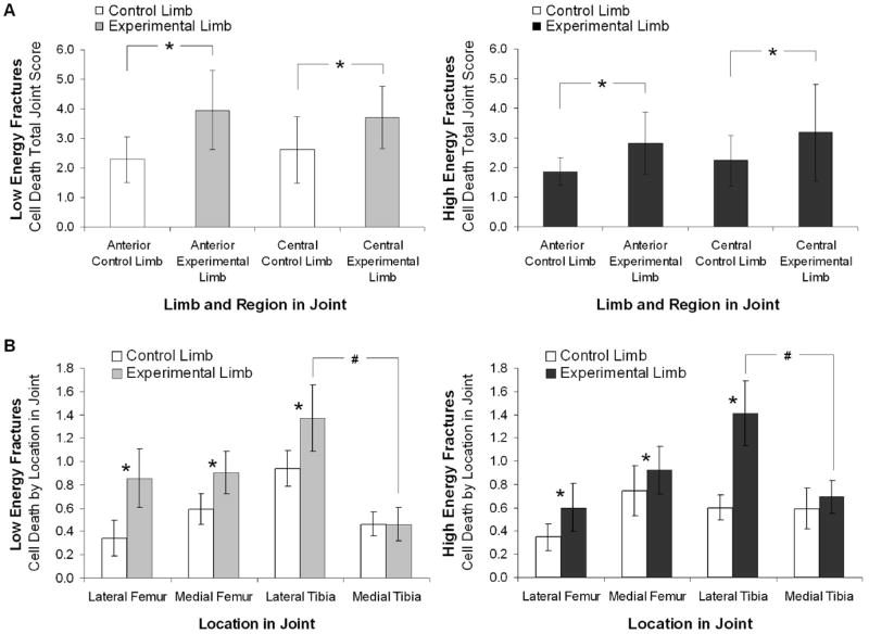
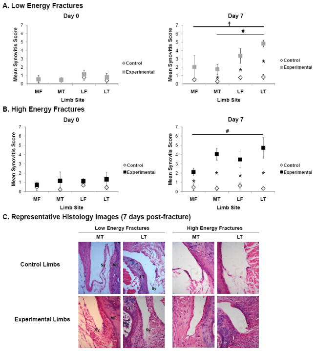
Design: Low and high energy articular fractures (n=25 per group) of the tibial plateau were created in adult male C57BL/6 mice. The acute effect of articular fracture severity on synovial inflammation, bone morphology, liberated fracture area, cartilage pathology, chondrocyte viability, and systemic cytokines and biomarkers levels was assessed at 0, 1, 3, 5, and 7 days post-fracture.
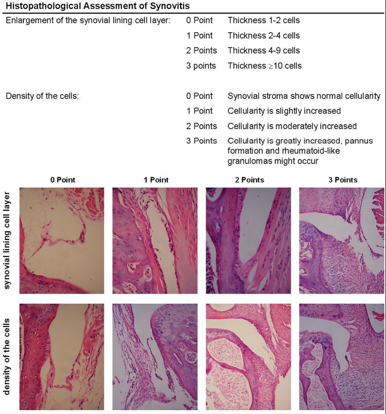
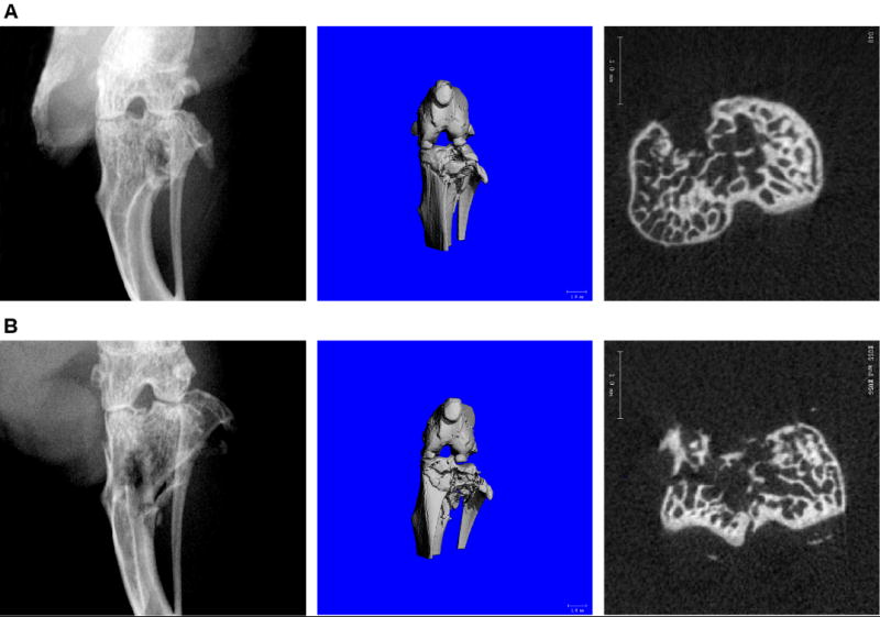
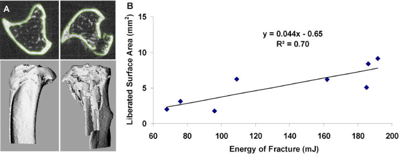
Results: Increasing intra-articular fracture severity was associated with greater acute pathology in the synovium and bone compared to control limbs, including increased global synovitis and reduced periarticular bone density and thickness. Applied fracture energy was significantly correlated with degree of liberated cortical bone surface area, indicating greater comminution. Serum concentrations of hyaluronic acid (HA) were significantly increased 1 day post-fracture. While articular fracture significantly reduced chondrocyte viability, there was no relationship between fracture severity and chondrocyte viability, cartilage degeneration, or systemic levels of cytokines and biomarkers.
Conclusions: This study demonstrates that articular fracture is associated with a loss of chondrocyte viability and increased levels of systemic biomarkers, and that increased intra-articular fracture severity is associated with increased acute joint pathology in a variety of joint tissues, including synovial inflammation, cortical comminution, and bone morphology. Further characterization of the early events following articular fracture could aid in the treatment of post-traumatic arthritis.
Copyright © 2011. Published by Elsevier Ltd.
Conflict of interest statement
Figures
References
-
- Swiontkowski MF, Chapman JR. Cost and effectiveness issues in care of injured patients. Clinical Orthopaedics & Related Research. 1995:17–24. - PubMed
-
- Brown TD, Johnston RC, Saltzman CL, Marsh JL, Buckwalter JA. Post-Traumatic Osteoarthritis: A First Estimate of Incidence, Prevalence, and Burden of Disease. Journal of Orthopaedic Trauma. 2006;20:739–44. - PubMed
-
- Swiontkowski MF, Agel J, McAndrew MP, Burgess AR, MacKenzie EJ. Outcome validation of the AO/OTA fracture classification system. J Orthop Trauma. 2000;14:534–41. - PubMed
-
- Lovasz G, Llinas A, Benya PD, Park SH, Sarmiento A, Luck JV., Jr Cartilage changes caused by a coronal surface stepoff in a rabbit model. Clin Orthop Relat Res. 1998:224–34. - PubMed
Publication types
MeSH terms
Substances
Grants and funding
LinkOut - more resources
Full Text Sources
Other Literature Sources
