. 2011 May 27;145(5):732-44.
doi: 10.1016/j.cell.2011.03.054.

Pyruvate kinase M2 is a PHD3-stimulated coactivator for hypoxia-inducible factor 1

Affiliations

Pyruvate kinase M2 is a PHD3-stimulated coactivator for hypoxia-inducible factor 1

Weibo Luo et al. Cell. .

Abstract

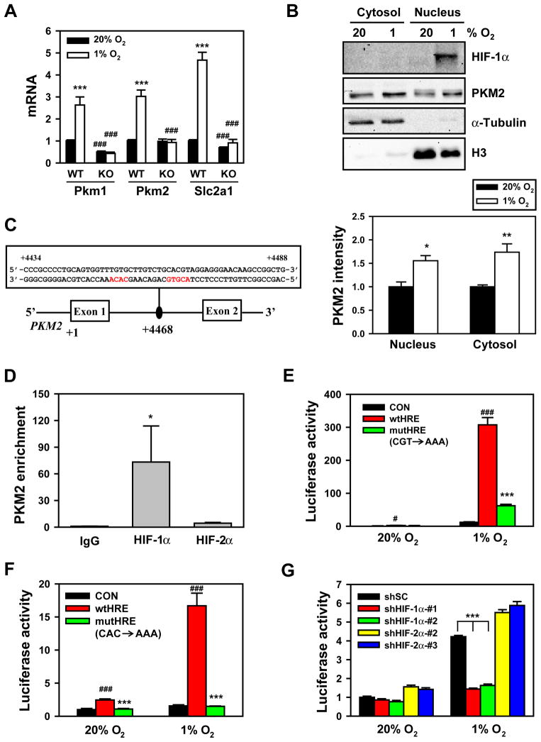
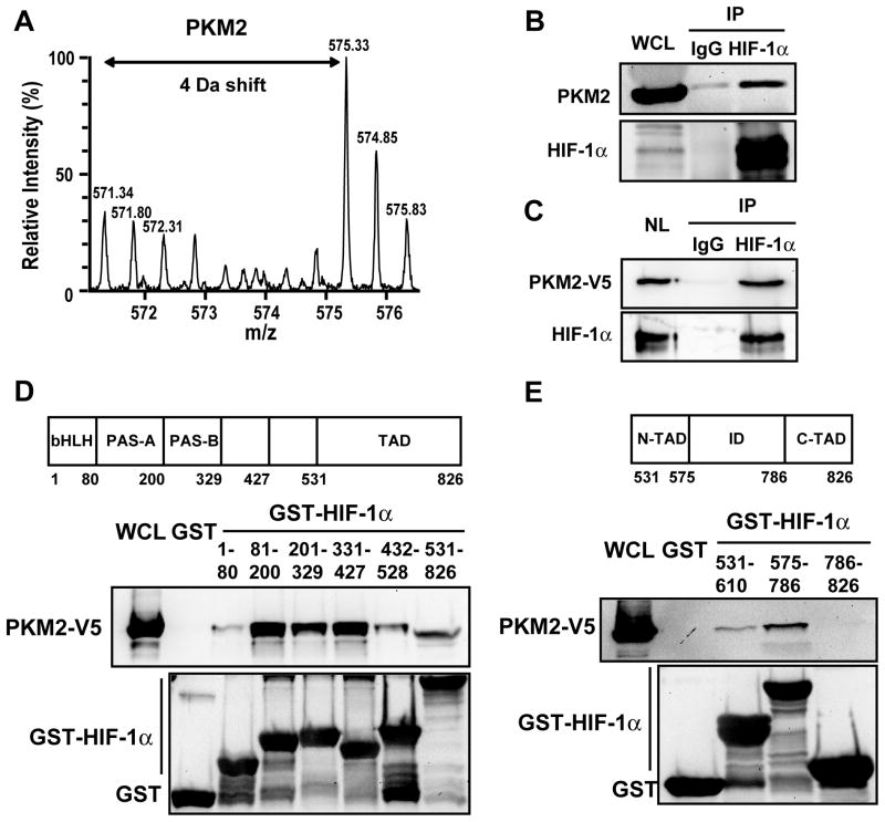
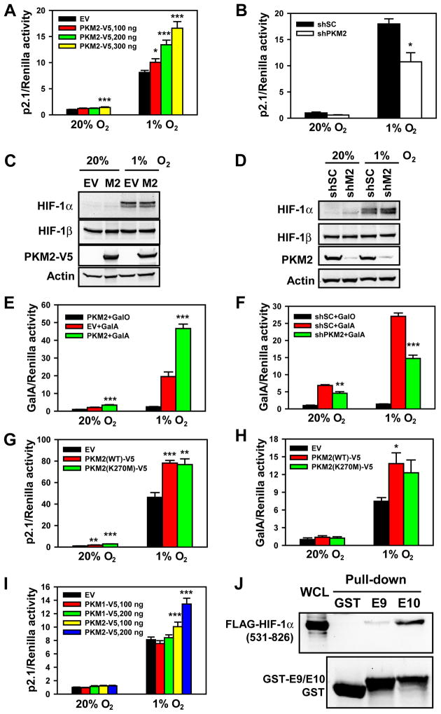
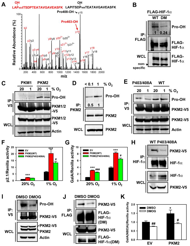
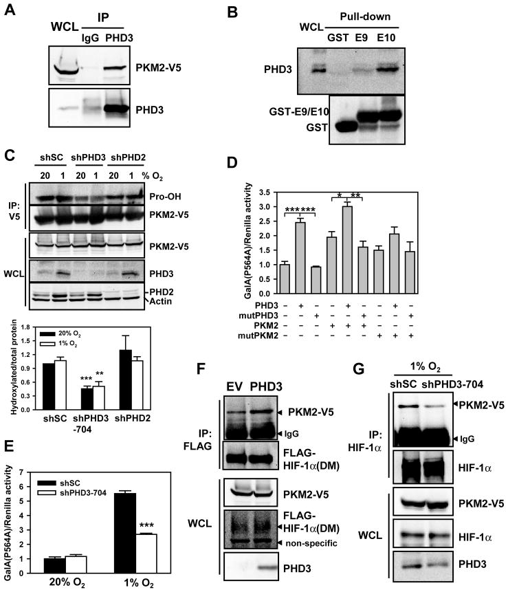
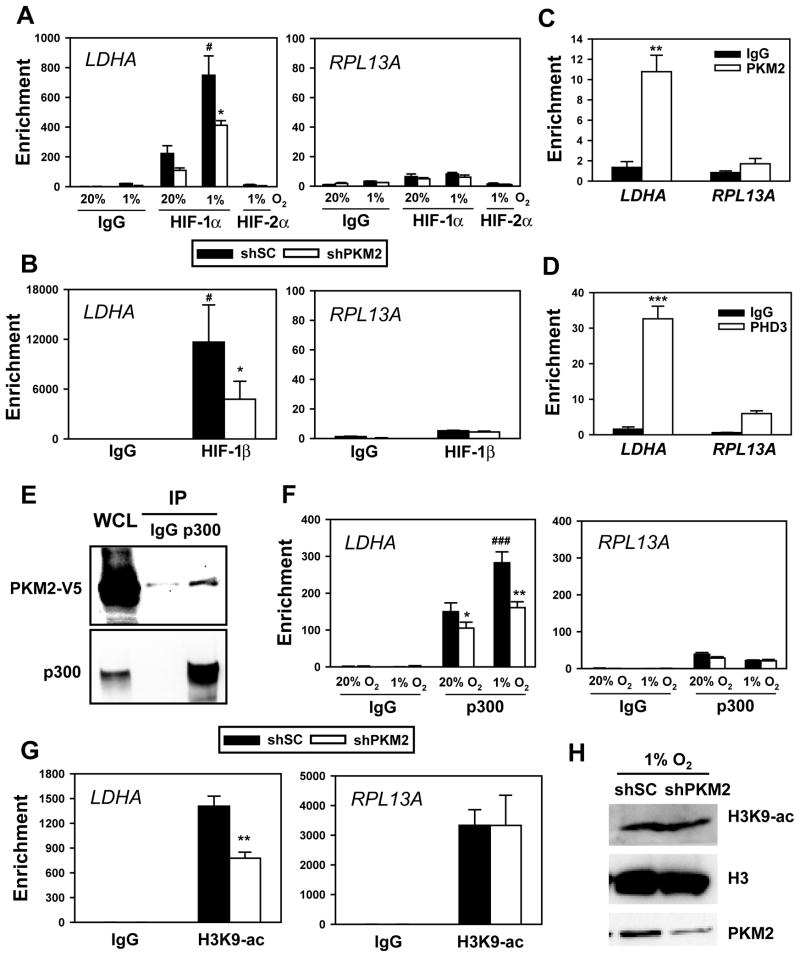
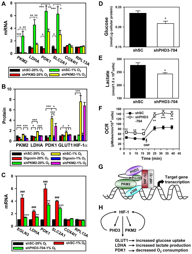
The pyruvate kinase isoforms PKM1 and PKM2 are alternatively spliced products of the PKM2 gene. PKM2, but not PKM1, alters glucose metabolism in cancer cells and contributes to tumorigenesis by mechanisms that are not explained by its known biochemical activity. We show that PKM2 gene transcription is activated by hypoxia-inducible factor 1 (HIF-1). PKM2 interacts directly with the HIF-1α subunit and promotes transactivation of HIF-1 target genes by enhancing HIF-1 binding and p300 recruitment to hypoxia response elements, whereas PKM1 fails to regulate HIF-1 activity. Interaction of PKM2 with prolyl hydroxylase 3 (PHD3) enhances PKM2 binding to HIF-1α and PKM2 coactivator function. Mass spectrometry and anti-hydroxyproline antibody assays demonstrate PKM2 hydroxylation on proline-403/408. PHD3 knockdown inhibits PKM2 coactivator function, reduces glucose uptake and lactate production, and increases O(2) consumption in cancer cells. Thus, PKM2 participates in a positive feedback loop that promotes HIF-1 transactivation and reprograms glucose metabolism in cancer cells.


Figures

Figure 1
Figure 1. HIF-1 Regulates Hypoxia-Induced Expression of PKM1 and PKM2
(A) qRT-PCR analysis of Pkm1, Pkm2 and Slc2a1 mRNA in wild-type (WT) and HIF-1α knockout (KO) mouse embryo fibroblasts (MEFs) exposed to 20% or 1% O2 for 24 h (mean ± SEM, n = 3–5). ***p<0.001 vs 20% O2. ###p<0.001 vs WT MEFs. (B) Immunoblot assays of HIF-1α, PKM2, histone H3, and α-tubulin protein in nuclear and cytosolic lysates prepared from HeLa cells exposed to 20% or 1% O2 for 24 h. The intensity of PKM2 was quantified by densitometry and normalized to 20% O2 (mean ± SEM, n = 3–4). *p<0.05, **p<0.01 vs 20% O2. (C) Nucleotide sequence of the hypoxia-response element (HIF-1 binding site 5′-ACGTG-3′ and 5′-CACA-3′ site shown in red) within intron 1 of the human PKM2 gene. The transcription initiation site is designated +1. Exons and intron are not drawn to scale. (D) Chromatin was precipitated from DMOG-treated HeLa cells (4 h) with antibodies against HIF-1α, HIF-2α, or IgG, and analyzed by qPCR (mean ± SEM, n = 4–5). *p<0.05 vs IgG. (E and F) HeLa cells were co-transfected with pGL2p control (CON), pGL2p-WT PKM2 HRE (wtHRE), or pGL2p-mutant PKM2 HRE (mutHRE) with CGT→AAA (E) or CAC→AAA (F), and pSV-Renilla, and exposed to 20% or 1% O2 for 24 h. The ratio of FLuc:RLuc activity was normalized to CON at 20% O2 (mean ± SEM, n = 4). #p<0.05, ###p<0.001 vs CON; ***p<0.001 vs wtHRE. (G) HeLa cells were transfected with: vector encoding a short hairpin (sh) RNA targeting HIF-1α, HIF-2α, or a scrambled control (SC); pGL2p-wtHRE; and pSV-Renilla, and exposed to 20% or 1% O2 for 24 h. The ratio of FLuc:RLuc activity was normalized to shSC at 20% O2 (mean ± SEM, n = 4). ***p<0.001 vs shSC. See also Figure S1.
Figure 2
Figure 2. PKM2 Interacts with HIF-1α
(A) GST pull-down assays were performed with GST or GST-HIF-1α (531–826) and lysate prepared from HEK293 cells grown in media containing light isotopes (12C6-14N2-Lys and 12C6-14N4-Arg) or heavy isotopes (13C6-15N2-Lys and 13C6-15N4-Arg), respectively. The precipitated proteins were analyzed by Q-TOF mass spectrometry (Luo et al., 2010). The mass spectrum of a tryptic PKM2 peptide is shown. Monoisotopic peaks derived from PKM2 bound to GST-HIF-1α (531–826) are 4 Da greater in their mass/charge ratio (m/z) and have higher relative intensity compared to the corresponding peaks due to non-specific binding of PKM2 to GST. (B) Co-IP was performed in HeLa cells exposed to 1% O2 for 24 h. (C) Co-IP was performed using nuclear lysates (NL) prepared from HeLa cells transfected with PKM2-V5 vector and exposed to 1% O2 for 24 h. (D and E) GST pull-down assays were performed with GST or GST fusion protein containing the indicated amino acid residues of HIF-1α (upper panels) and whole cell lysates (WCLs) from HeLa cells expressing PKM2-V5. bHLH, basic helix-loop-helix; PAS, Per-ARNT-Sim; TAD, transactivation domain; ID, inhibitory domain.
Figure 3
Figure 3. PKM2 Promotes HIF-1 Transactivation
(A, B, G and I) HeLa cells were transfected with p2.1, pSV-Renilla, and empty vector (EV) or the indicated expression vector, and exposed to 20% or 1% O2 for 24 h. The ratio of FLuc:RLuc activity was normalized to EV (A, G and I) or shSC (B) at 20% O2 (mean ± SEM, n = 4). *p<0.05, **p<0.01, ***p<0.001 vs EV or shSC. (C) Immunoblot assays of HIF-1α, HIF-1β, PKM2-V5, and actin in HeLa cells transfected with EV or PKM2-V5 (M2) expression vector and exposed to 20% or 1% O2 for 4 h. (D) HeLa cells were transduced with a retrovirus encoding shSC or two retroviruses encoding different shRNAs targeting PKM2 (shM2), exposed to 20% or 1% O2 for 4 h, and WCLs were subjected to immunoblot assays. (E, F and H) HeLa cells were transfected with GalA or GalO, pG5E1bLuc, and pSV-Renilla, and the indicated expression vector, and exposed to 20% or 1% O2 for 24 h. The ratio of FLuc:RLuc activity was normalized to GalO (E and F) or EV (H) at 20% O2 (mean ± SEM, n = 4). *p<0.05, **p<0.01, ***p<0.001 vs EV or shSC. (J) GST pull-down assays were performed with GST, GST-E9, or GST-E10 and WCLs from HeLa cells expressing FLAG-HIF-1α (531–826). See also Figure S2.
Figure 4
Figure 4. Prolyl Hydroxylation of PKM2 Stimulates HIF-1 Transactivation
(A) GST-E10 was incubated with WCLs to induce hydroxylation and tryptic peptides were analyzed by mass spectrometry. The fragmentation spectrum of 401LAPITSDPTEATAVGAVEASFK422 revealed the presence of peptides with hydroxylation of Pro-403 or Pro-408. (B) HEK293 cells were transfected with WT FLAG-HIF-1α or double mutant (DM) FLAG-HIF-1α(P402A/P564A) and treated with the proteasome inhibitor MG132 (10 μM) for 4 h. IP was performed with anti-FLAG antibody, followed by immunoblot assay. The intensity of hydroxylated FLAG-HIF-1α was quantified by densitometry and normalized to WT. (C) HeLa cells were transfected with vector encoding PKM2-V5 or PKM1-V5 and exposed to 20% or 1% O2 for 24 h. IP was performed with anti-V5 agarose, followed by immunoblot assay. (D) HeLa cells were exposed to 1% or <0.1% O2 for 4 h. IP was performed with anti-PKM2 antibody, followed by immunoblot assay. The intensity of hydroxylated PKM2 was quantified by densitometry and normalized to 1% O2. (E) HeLa cells were transfected with vector encoding WT PKM2-V5 or PKM2(P403/408A)-V5 and exposed to 20% or 1% O2 for 24 h. IP was performed with anti-V5 agarose, followed by immunoblot assay. (F and G) HeLa cells were co-transfected with p2.1 (F) or GalA(P564A) and pG5E1bLuc (G); pSV-Renilla; and the indicated expression vector, and exposed to 20% or 1% O2 for 24 h. The ratio of FLuc:RLuc activity was normalized to EV at 20% O2 (mean ± SEM, n = 4). ***p<0.001 vs EV; #p<0.05, ###p<0.001 vs PKM2(WT). (H) Co-IP was performed in HeLa cells transfected with vector encoding WT PKM2-V5 or PKM2(P403/408A)-V5 and exposed to 1% O2 for 4 h. (I) HeLa cells were transfected with PKM2-V5 vector and treated with DMSO or DMOG (100 μM) for 4 h. IP was performed with anti-V5 agarose, followed by immunoblot assay. (J) Co-IP was performed in HeLa cells co-transfected with FLAG-HIF-1α(DM) and PKM2-V5 vectors and treated with DMSO or DMOG for 4 h. (K) HeLa cells were co-transfected with GalA(N803A) vector, pG5E1bLuc, pSV-Renilla, and EV or PKM2-V5 vector, and treated with DMSO or DMOG for 24 h. The ratio of FLuc:RLuc activity was normalized to EV + DMSO (mean ± SEM, n = 4). *p<0.05 vs EV; #p<0.05, ##p<0.01 vs DMSO. See also Figure S3.
Figure 5
Figure 5. PHD3 Stimulates the Coactivator Function of PKM2
(A) Co-IP was performed in HeLa cells co-transfected with PKM2-V5 and PHD3 vectors. (B) GST pull-down assays were performed with purified GST, GST-E9, or GST-E10 and WCLs from HeLa cells exposed to 1% O2 for 24 h. (C) HeLa-shSC, HeLa-shPHD3-704, and HeLa-shPHD2 cells were transfected with PKM2-V5 vector and exposed to 20% or 1% O2 for 24 h. IP was performed with anti-V5 agarose, followed by immunoblot assay. Hydroxylated PKM2-V5 was quantified by densitometry and normalized to total immunoprecipitated PKM2-V5 (mean ± SEM, n = 3). **p<0.01, ***p<0.001 vs shSC. (D) RCC4 cells were co-transfected with GalA(P564A) vector, pG5E1bLuc, pSV-Renilla, and the indicated expression vector for 24 h. The ratio of FLuc:RLuc activity was normalized to EV (mean ± SEM, n = 4). *p<0.05; **p<0.01; ***p<0.001. mutPHD3, PHD3(H135A/D137A); mutPKM2, PKM2(P403/408A). (E) HeLa cells were co-transfected with GalA(P564A) vector, pG5E1bLuc, and pSV-Renilla, and the indicated expression vector and exposed to 20% or 1% O2 for 24 h. The ratio of FLuc:RLuc activity was normalized to shSC at 20% O2 (mean ± SEM, n = 4). ***p<0.001 vs shSC. (F) Co-IP was performed in HeLa cells co-transfected with vector encoding double mutant (DM) FLAG-HIF-1α(P402A/P564A), EV or PHD3, and PKM2-V5. (G) Co-IP was performed in HeLa cells co-transfected with vectors encoding PKM2-V5 and either shSC or shPHD3 and exposed to 1% O2 for 24 h. See also Figure S4.
Figure 6
Figure 6. PKM2 Enhances HIF-1 Binding and p300 Recruitment to HREs
(A) HeLa cells were transfected with shSC or shPKM2-#1 vector and exposed to 20% or 1% O2 for 4 h. ChIP assays were performed with IgG, anti-HIF-1α, or anti-HIF-2α antibody (mean ± SEM, n = 4). #p<0.05 vs shSC-20% O2; *p<0.05 vs shSC-1% O2. (B) HeLa cells were transfected with shSC or shPKM2-#1 vector, and exposed to 1% O2 for 4 h. ChIP assays were performed with IgG or anti-HIF-1β antibody (mean ± SEM, n = 3). #p<0.05 vs IgG; *p<0.05 vs shSC. (C and D) HeLa cells were exposed to 1% O2 for 24 h. ChIP assays were performed with IgG, anti-PKM2 (C), or anti-PHD3 (D) antibody (mean ± SEM, n = 3). **p<0.01, ***p<0.001 vs IgG. (E) Co-IP was performed in HeLa cells transfected with PKM2-V5 vector for 24 h. (F) HeLa cells expressing shSC or shPKM2-#1 were exposed to 20% or 1% O2 for 24 h. ChIP assays were performed with IgG or anti-p300 antibody (mean ± SEM, n = 3). *p<0.05, **p<0.01 vs shSC; ###p<0.001 vs shSC-20% O2. (G) HeLa cells were transfected with shSC or shPKM2-#1 vector and exposed to 1% O2 for 24 h. ChIP assays were performed with IgG or anti-acetylated H3K9 (H3K9-ac) antibody (mean ± SEM, n = 3). **p<0.01 vs shSC. (H) Immunoblot assays of total H3K9-ac, histone H3, and PKM2 in HeLa cells transfected with shSC or shPKM2-#1 vector and exposed to 1% O2 for 24 h. See also Figure S5.
Figure 7
Figure 7. PKM2 and PHD3 Promote HIF-1-dependent Glycolytic Metabolism
(A) qRT-PCR analysis of indicated mRNAs in HeLa cells transfected with shSC or shPKM2-#1 vector and exposed to 20% or 1% O2 for 24 h (mean ± SEM, n = 4). *p<0.05, **p<0.01, ***p<0.001. (B) HeLa cells were transduced with shSC or shPKM2-#1 and shPKM2-#2 retrovirus, and exposed to 20% or 1% O2 in the absence or presence of digoxin (100 nM) for 24 h. Levels of the indicated protein were determined by immunoblot assay, quantified by densitometry, and normalized to actin (mean ± SEM, n = 4). *p<0.05, ***p<0.001. (C) qRT-PCR analysis of indicated mRNAs in HeLa cells transduced with shSC or shPHD3-704 retrovirus and exposed to 20% or 1% O2 for 24 h (mean ± SEM, n = 3–4). ###p<0.001 vs shSC-20% O2; **p<0.01, ***p<0.001 vs shSC-1% O2. (D–F) RCC4 cells were transduced with shSC or shPHD3-704 retrovirus. Glucose was measured in lysates and normalized to total cellular protein amount (mean ± SEM, n = 3; D); lactate was measured in the culture media and normalized to cell number (mean ± SEM, n = 3; E); O2 consumption rate (OCR) was measured and normalized to cell number (mean ± SEM, n = 4–8; F). *p<0.05, **p<0.01, ***p<0.01 vs shSC. (G) PKM2 is prolyl hydroxylated by PHD3 and interacts with HIF-1α and p300, thereby enhancing HRE occupancy by HIF-1 and p300, and increasing histone acetylation. (H) Feed-forward mechanism for HIF-1 activity. HIF-1 activates transcription of genes encoding PHD3 and PKM2, which interact with HIF-1α to stimulate transactivation of target genes encoding GLUT1, LDHA, PDK1, and other metabolic enzymes that mediate the Warburg effect in cancer cells. See also Figure S6.

Comment in

References

    1. Arany Z, Huang LE, Eckner R, Bhattacharya S, Jiang C, Goldberg MA, Bunn HF, Livingston DM. An essential role for p300/CBP in the cellular response to hypoxia. Proc Natl Acad Sci U S A. 1996;93:12969–12973. - PMC - PubMed
    1. Berra E, Benizri E, Ginouves A, Volmat V, Roux D, Pouyssegur J. HIF prolyl-hydroxylase 2 is the key oxygen sensor setting low steady-state levels of HIF-1α in normoxia. EMBO J. 2003;22:4082–4090. - PMC - PubMed
    1. Christofk HR, Vander Heiden MG, Harris MH, Ramanathan A, Gerszten RE, Wei R, Fleming MD, Schreiber SL, Cantley LC. The M2 splice isoform of pyruvate kinase is important for cancer metabolism and tumour growth. Nature. 2008;452:230–233. - PubMed
    1. David CJ, Chen M, Assanah M, Canoll P, Manley JL. HnRNP proteins controlled by c-Myc deregulate pyruvate kinase mRNA splicing in cancer. Nature. 2010;463:364–368. - PMC - PubMed
    1. Dombrauckas JD, Santarsiero BD, Mesecar AD. Structural basis for tumor pyruvate kinase M2 allosteric regulation and catalysis. Biochemistry. 2005;44:9417–9429. - PubMed

Publication types

MeSH terms

Substances