. 2011 Jun 28;58(1):73-82.
doi: 10.1016/j.jacc.2010.12.044. Epub 2011 May 27.

Mitochondrial targeted antioxidant Peptide ameliorates hypertensive cardiomyopathy

Affiliations

Mitochondrial targeted antioxidant Peptide ameliorates hypertensive cardiomyopathy

Dao-Fu Dai et al. J Am Coll Cardiol. .

Abstract

Objectives: We investigated the effect of reducing mitochondrial oxidative stress by the mitochondrial-targeted antioxidant peptide SS-31 in hypertensive cardiomyopathy.

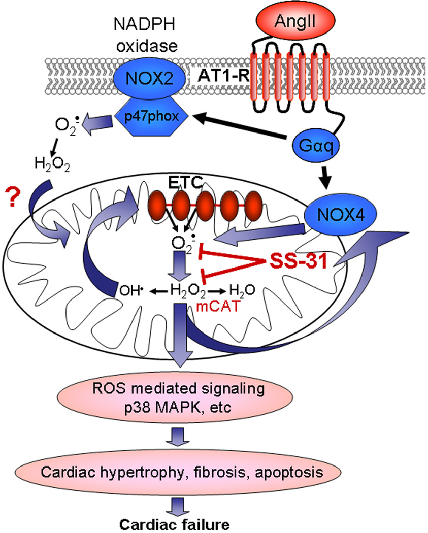
Background: Oxidative stress has been implicated in hypertensive cardiovascular diseases. Mitochondria and nicotinamide adenine dinucleotide phosphate (NADPH) oxidase have been proposed as primary sites of reactive oxygen species (ROS) generation.

Methods: The mitochondrial targeted antioxidant peptide SS-31 was used to determine the role of mitochondrial oxidative stress in angiotensin II (Ang)-induced cardiomyopathy as well as in Gαq overexpressing mice with heart failure.

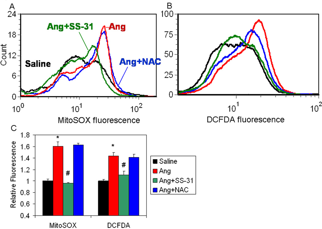
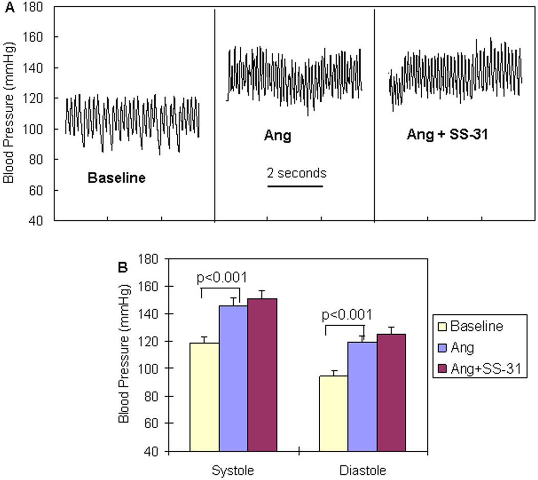
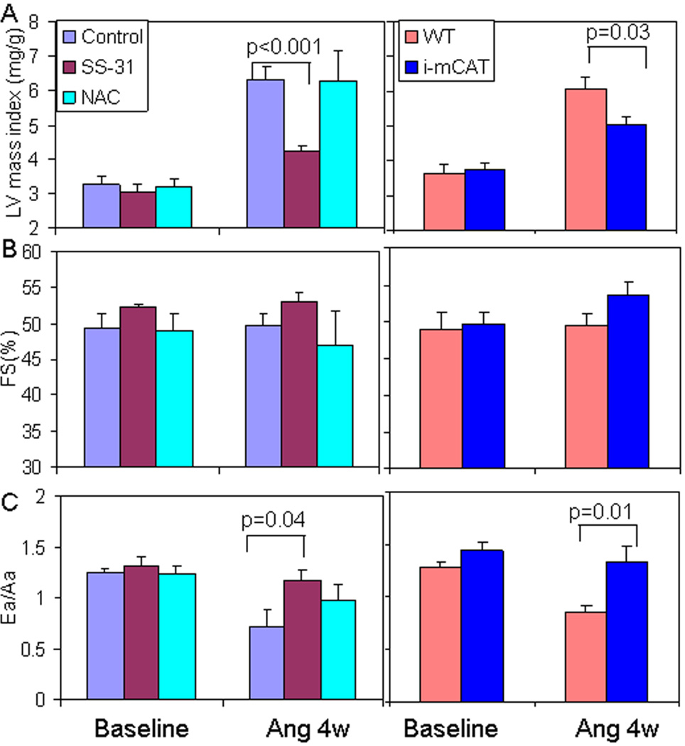
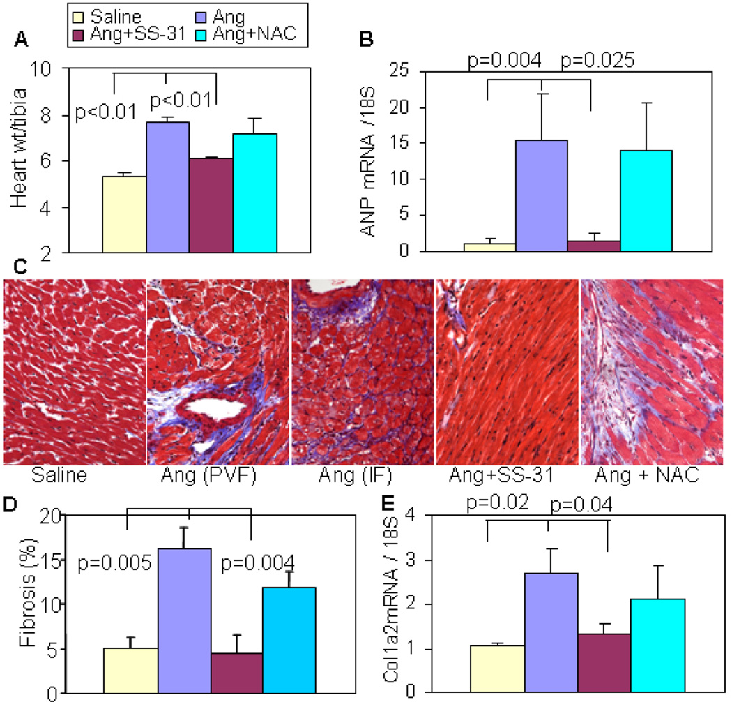
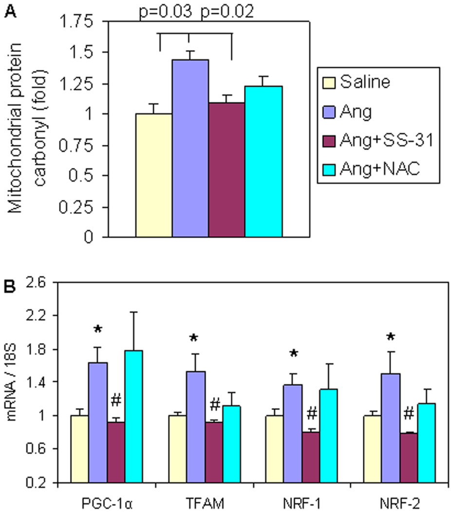
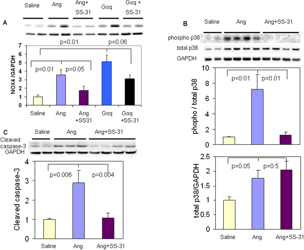
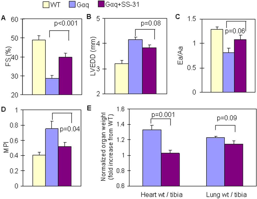
Results: Ang induces mitochondrial ROS in neonatal cardiomyocytes, which is prevented by SS-31, but not the nontargeted antioxidant N-acetyl cysteine (NAC). Continuous administration of Ang for 4 weeks in mice significantly increased both systolic and diastolic blood pressure, and this was not affected by SS-31 treatment. Ang was associated with up-regulation of NADPH oxidase 4 (NOX4) expression and increased cardiac mitochondrial protein oxidative damage, and induced the signaling for mitochondrial biogenesis. Reducing mitochondrial ROS by SS-31 substantially attenuated Ang-induced NOX4 up-regulation, mitochondrial oxidative damage, up-regulation of mitochondrial biogenesis, and phosphorylation of p38 mitogen-activated protein kinase and prevented apoptosis, concomitant with amelioration of Ang-induced cardiac hypertrophy, diastolic dysfunction, and fibrosis, despite the absence of blood pressure-lowering effect. The NAC did not show any beneficial effect. The SS-31 administration for 4 weeks also partially rescued the heart failure phenotype of Gαq overexpressing mice.

Conclusions: Mitochondrial targeted peptide SS-31 ameliorates cardiomyopathy resulting from prolonged Ang stimulation as well as Gαq overexpression, suggesting its potential clinical application for target organ protection in hypertensive cardiovascular diseases.


Figures

Figure 1
Figure 1. Ang increased mitochondrial and total cellular oxidative stress in neonatal cardiomyocytes is prevented by SS-31 but not N-acetyl cysteine
Flow cytometry of neonatal cardiomyocytes stimulated with Ang (1µM) and loaded with MitoSOX (A) or CM-DCFDA (B). (C) Quantitative analysis (presented as mean±SEM of histogram medians, n=3–5) revealed a significant increase in MitoSOX and DCFDA fluorescence in Ang-treated cardiomyotyes (red), compared with saline treatment (black). The non-targeted antioxidant NAC (0.5mM, blue line) had no significant effect on Ang induced mitochondrial ROS (A, C) and total cellular ROS (B, C). Simultaneous treatment with SS-31 (1nM, green line) substantially reduced Ang induced mitochondrial ROS (A, C) and total cellular ROS (B, C).*p<0.01 vs. saline; #p<0.01 vs. Ang.
Figure 2
Figure 2. No effect of SS-31 on blood pressure increase after pressor dose of Ang
(A) Representative blood pressure tracings of mice at baseline and after Ang (1.1mg/kg/d) administered with a subcutaneous pump. (B) Ang significantly increased systolic and diastolic BP. Administration of SS-31 (3mg/kg/d) did not show any significant effect on BP when added to Ang treatment. (n=3)
Figure 3
Figure 3. SS-31 ameliorates Ang-induced cardiac hypertrophy and diastolic dysfunction
(A) Ang for 4 weeks substantially increased LV mass index in control mice. Simultaneous administration of SS-31 significantly attenuated this increase in LVMI (left panel). This was similar extent to that observed in mice with inducible overexpression of mitochondrial catalase (i-mCAT, right panel). (B) Fractional shortening (FS,%) was not significantly changed after 4 weeks of Ang in the presence or absence of mitochondrial antioxidants. (C) Diastolic function measured by tissue Doppler imaging of Ea/Aa was significantly reduced after Ang, but was significantly ameliorated by SS-31 or genetic overexpression of mCAT. Administration of NAC for 4 weeks did not confer any significant protection for Ang-induced hypertrophy and diastolic dysfunction (light blue bar on left panel, A–C). n=6–7
Figure 4
Figure 4. Ang-induced cardiac hypertrophy and fibrosis were attenuated by SS-31 but not oral NAC
(A) Ang significantly increased heart weight (normalized to tibia length), and this was significantly attenuated by SS-31 but not by NAC. (B) qPCR showed a dramatic increase in ANP gene expression, which was significantly prevented by SS-31, but not by NAC. (C) Representative histopathology showing that Ang induced substantial perivascular fibrosis (PVF) and interstitial fibrosis (IF), which was protected by SS-31 but not NAC. (D) Analysis of blue trichrome staining demonstrated a significant increase in ventricular fibrosis after Ang, and this was substantially attenuated by SS-31 but not NAC. (E). qPCR showed upregulation of pro-collagen1a2 mRNA after Ang, which was significantly reduced in SS-31 hearts but not in NAC hearts. n=4–7.
Figure 5
Figure 5. Mitochondrial protein carbonyl and signaling for mitochondrial biogenesis increased after 4 weeks of Ang treatment, which was prevented by SS-31
(A) Ang significantly increased cardiac mitochondrial protein carbonyl content, and was significantly ameliorated by SS-31, but not by NAC. (B) qPCR revealed significant upregulation of genes in mitochondrial biogenesis, all of which were attenuated by SS-31. NAC treatment did not significantly change any genes in mitochondrial biogenesis signaling. *p–0.05 compared with saline group, #p–0.05 compared with Ang treated group, n=4–7.
Figure 6
Figure 6. SS-31 reduces Ang-induced upregulation of NOX4, activation of p38 MAPK and apoptosis
(A) NADPH oxidase-4 (NOX4) protein was significantly increased after 4 weeks of Ang or by Gαq overexpression, and this was significantly attenuated by SS-31. (B) Ang for 4 weeks substantially induced apoptosis, as shown by increases cleaved caspase 3 message, and this was significantly attenuated by SS-31. (C) Phosphorylation of p38 MAP kinase was significantly increased after Ang, but was substantially lower in SS-31 treated hearts (upper panel). Protein levels of p38 MAP kinase also increased after Ang, n=3–6.
Figure 7
Figure 7. SS-31 ameliorated cardiac hypertrophy and failure in Gαq overexpressing mice
Echocardiography of Gαq mice 16 weeks of age. (A) SS-31 (3mg/kg/d) for 4 weeks (from age 12–16 weeks) significantly ameliorated the decline in systolic function, as indicated by FS in Gαq mice. (B,C) Chamber enlargement and diastolic dysfunction in Gαq mice were slightly attenuated by SS-31 with a statistical trend, p=0.08 and 0.06, respectively. (D) Worsening of myocardial performance index (MPI) in Gαq mice was significantly ameliorated by SS-31. (E) An increase in normalized heart weight in Gαq mice was substantially protected by SS-31, while there was a trend towards protection from increased normalized lung weight by SS-31 (p=0.09), n=3–6.
Figure 8
Figure 8. Diagrammatic illustration of the proposed mechanisms of SS-31 protection in Ang- and Gαq-induced cardiomyopathy
Ang and Gαq activate NOX isoforms that may directly (NOX4) or indirectly (NOX2) elevate mitochondrial ROS. Amplification by ROS induced ROS mechanisms may facilitate ROS-mediated activation of signaling in hypertrophy and failure.

Comment in

References

    1. Lawes CM, Vander Hoorn S, Rodgers A. Global burden of blood-pressure-related disease, 2001. Lancet. 2008;371:1513–1518. - PubMed
    1. Gardin JM, Lauer MS. Left ventricular hypertrophy: the next treatable, silent killer? Jama. 2004;292:2396–2398. - PubMed
    1. Mollnau H, Wendt M, Szocs K, et al. Effects of angiotensin II infusion on the expression and function of NAD(P)H oxidase and components of nitric oxide/cGMP signaling. Circ Res. 2002;90:E58–E65. - PubMed
    1. Kimura S, Zhang GX, Nishiyama A, et al. Mitochondria-derived reactive oxygen species and vascular MAP kinases: comparison of angiotensin II and diazoxide. Hypertension. 2005;45:438–444. - PubMed
    1. Doughan AK, Harrison DG, Dikalov SI. Molecular mechanisms of angiotensin IImediated mitochondrial dysfunction: linking mitochondrial oxidative damage and vascular endothelial dysfunction. Circ Res. 2008;102:488–496. - PubMed

Publication types