. 2011 Sep;30(5):359-94.
doi: 10.1016/j.preteyeres.2011.05.001. Epub 2011 May 19.

Acute retinal arterial occlusive disorders

Affiliations

Acute retinal arterial occlusive disorders

Sohan Singh Hayreh. Prog Retin Eye Res. 2011 Sep.

Abstract

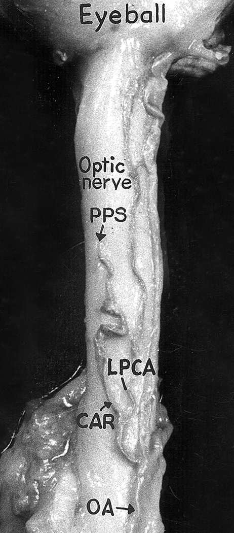
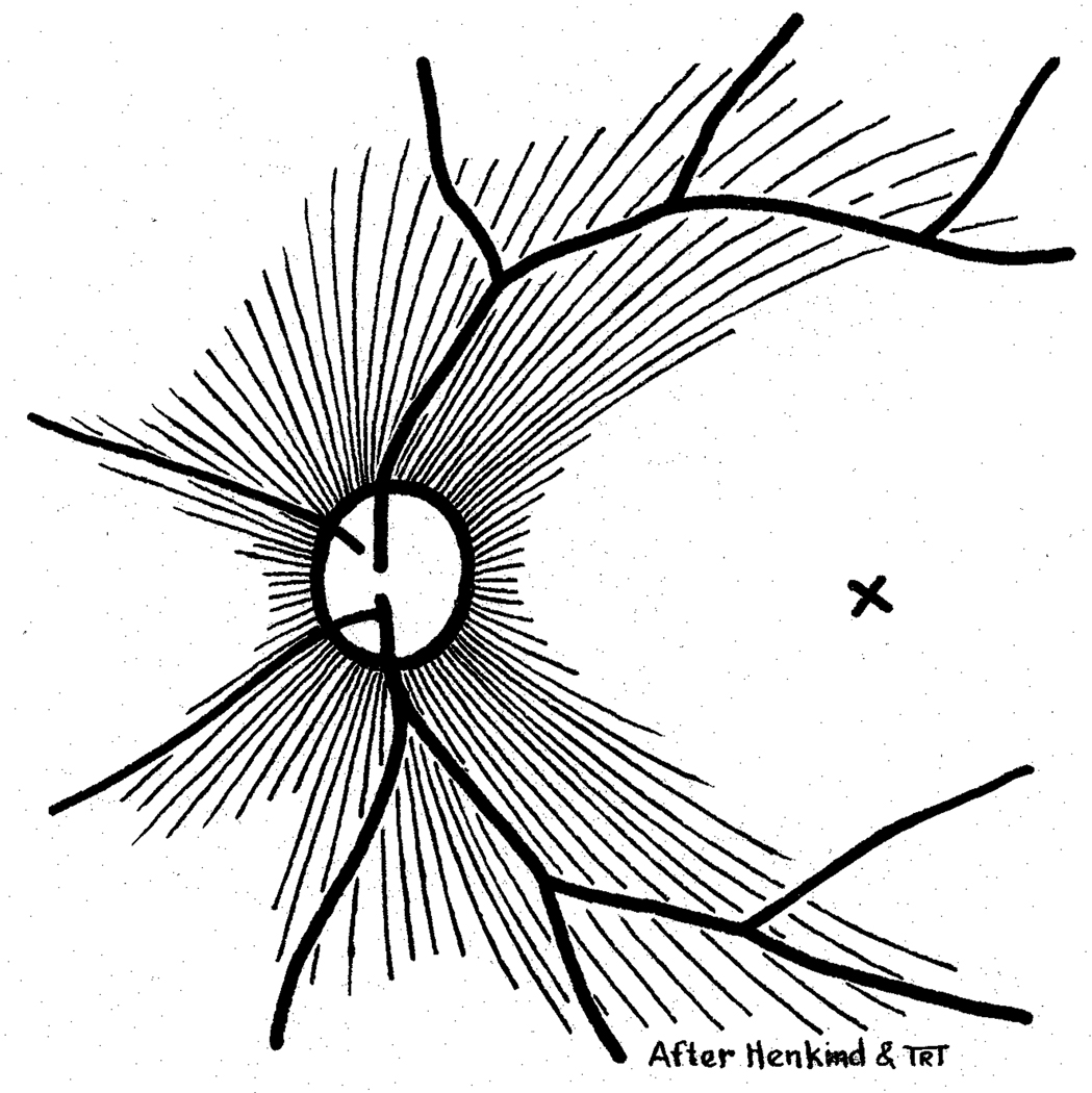
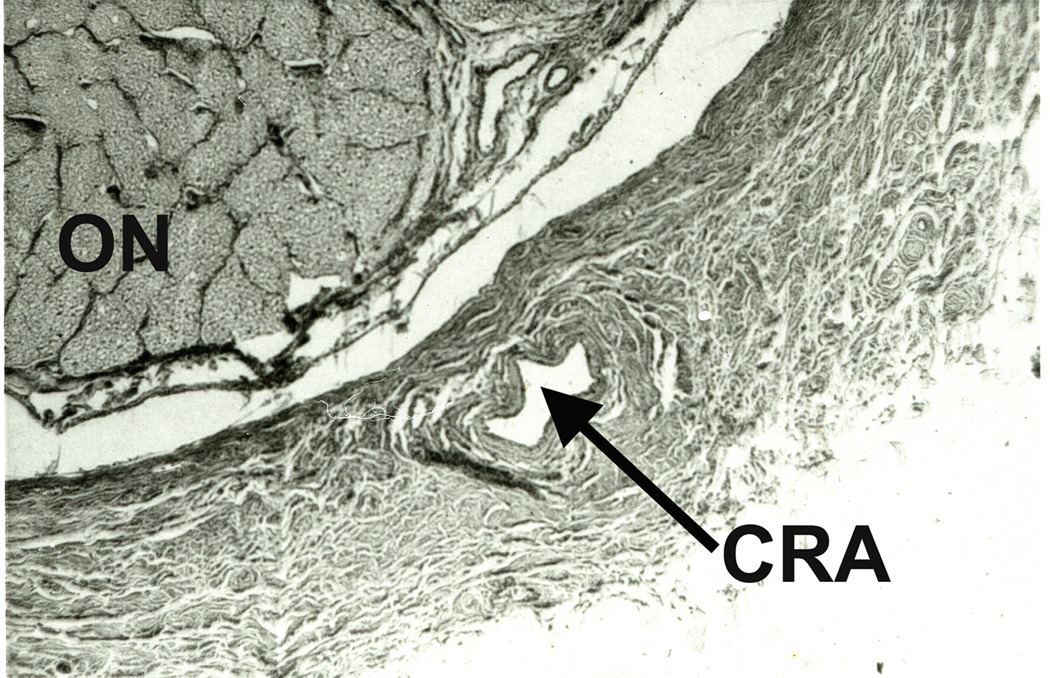
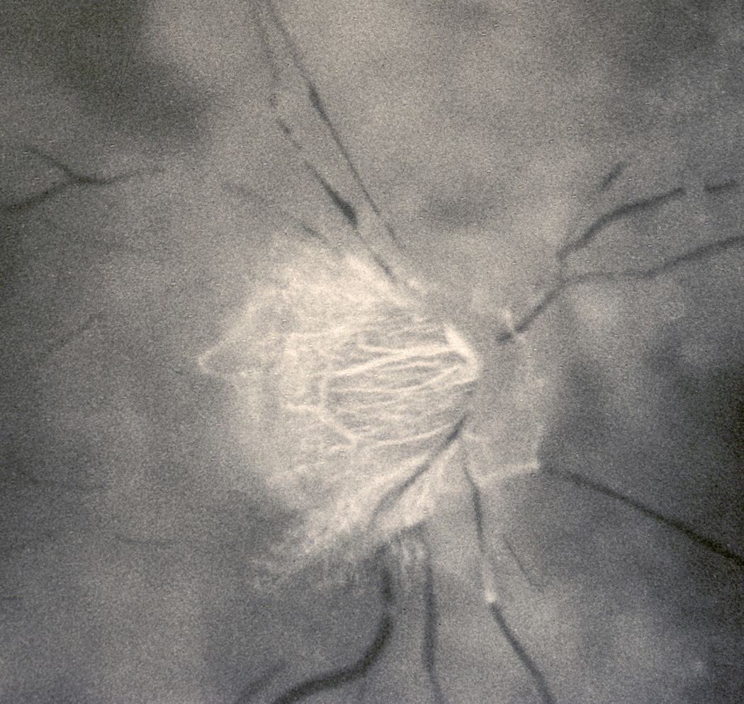
The initial section deals with basic sciences; among the various topics briefly discussed are the anatomical features of ophthalmic, central retinal and cilioretinal arteries which may play a role in acute retinal arterial ischemic disorders. Crucial information required in the management of central retinal artery occlusion (CRAO) is the length of time the retina can survive following that. An experimental study shows that CRAO for 97min produces no detectable permanent retinal damage but there is a progressive ischemic damage thereafter, and by 4h the retina has suffered irreversible damage. In the clinical section, I discuss at length various controversies on acute retinal arterial ischemic disorders. Classification of acute retinal arterial ischemic disorders: These are of 4 types: CRAO, branch retinal artery occlusion (BRAO), cotton wool spots and amaurosis fugax. Both CRAO and BRAO further comprise multiple clinical entities. Contrary to the universal belief, pathogenetically, clinically and for management, CRAO is not one clinical entity but 4 distinct clinical entities - non-arteritic CRAO, non-arteritic CRAO with cilioretinal artery sparing, arteritic CRAO associated with giant cell arteritis (GCA) and transient non-arteritic CRAO. Similarly, BRAO comprises permanent BRAO, transient BRAO and cilioretinal artery occlusion (CLRAO), and the latter further consists of 3 distinct clinical entities - non-arteritic CLRAO alone, non-arteritic CLRAO associated with central retinal vein occlusion and arteritic CLRAO associated with GCA. Understanding these classifications is essential to comprehend fully various aspects of these disorders. Central retinal artery occlusion: The pathogeneses, clinical features and management of the various types of CRAO are discussed in detail. Contrary to the prevalent belief, spontaneous improvement in both visual acuity and visual fields does occur, mainly during the first 7 days. The incidence of spontaneous visual acuity improvement during the first 7 days differs significantly (p<0.001) among the 4 types of CRAO; among them, in eyes with initial visual acuity of counting finger or worse, visual acuity improved, remained stable or deteriorated in non-arteritic CRAO in 22%, 66% and 12% respectively; in non-arteritic CRAO with cilioretinal artery sparing in 67%, 33% and none respectively; and in transient non-arteritic CRAO in 82%, 18% and none respectively. Arteritic CRAO shows no change. Recent studies have shown that administration of local intra-arterial thrombolytic agent not only has no beneficial effect but also can be harmful. Prevalent multiple misconceptions on CRAO are discussed. Branch retinal artery occlusion: Pathogeneses, clinical features and management of various types of BRAO are discussed at length. The natural history of visual acuity outcome shows a final visual acuity of 20/40 or better in 89% of permanent BRAO cases, 100% of transient BRAO and 100% of non-arteritic CLRAO alone. Cotton wools spots: These are common, non-specific acute focal retinal ischemic lesions, seen in many retinopathies. Their pathogenesis and clinical features are discussed in detail. Amaurosis fugax: Its pathogenesis, clinical features and management are described.


Conflict of interest statement

The author has no conflict of interest.

Figures

Figure 1
Figure 1
Variations in origin and course of the ophthalmic artery. A = Normal pattern; B,C,D, E = The ophthalmic artery arises firm the internal carotid artery as usual, but the major contribution comes from the middle meningeal artery. F,G = The only sources is the middle meningeal artery, as its connection with the internal carotid artery is either absent (F) or obliterated (G). Abbreviations: ICA = Internal carotid artery; Lac. = Lacrimal artery; MMA = Middle meningeal artery; OA = Ophthalmic artery. (Reproduced from Singh (Hayreh) et al. 1960a.)
Figure 2
Figure 2
Two trunks (1,2) of the central retinal artery, arising independently from the ophthalmic artery, as seen on splitting open the optic nerve (ON) behind the eyeball. (Reproduced from Hayreh 1958)
Figure 3
Figure 3
The central artery of the retina (CAR) arising by a common trunk with lateral posterior ciliary artery (LPCA) from the ophthalmic artery (OA). PPS = Point of penetration of the central artery of the retina into the optic nerve sheath. (Reproduced from Hayreh 1958)
Figure 4
Figure 4
Two views of the same optic nerve in the retrobulbar regions – (A) from inferior aspect and (B) from the superior aspect after removal of dural sheath. They show pial branches of the central retinal artery (CRA) arising from the intravaginal part of the artery, and their anastomoses anteriorly with recurrent pial branches of the circle of Zinn and Haller (CZ) and posteriorly with collateral (Col.) branches from the ophthalmic artery (O.A.). (Reproduced from Hayreh 1958)
Figure 5
Figure 5
This shows intravaginal (indicated by smaller arrow) and intraneural (anterior to the bigger arrow with asterisk where the optic nerve is split open to show the central region of the optic nerve) course of the central retinal artery (CRA) from below. It shows 2 pial branches (at and before the arrow with asterisk) and 6 intraneural branches anterior to that. (Reproduced from Hayreh 1958)
Figure 6
Figure 6
Schematic representation, showing: (A) The course of central retinal artery and its branches and anastomoses, central retinal vein and its tributaries, and blood supply of the optic nerve. (B) Blood vessels on the optic disc and in the retina. (Modified from Trans Am Acad Ophthalmol Otolaryngol 1974;78:OP240–OP254.). Abbreviations: A = arachnoid; C = choroid; CRA = central retinal artery; Col. Br. = Collateral branches; CRV = central retinal vein; D = dura; LC = lamina cribrosa; OD = optic disc; ON = optic nerve; PCA = posterior ciliary artery; PR = prelaminar region; R = retina; S = sclera; SAS = subarachnoid space.
Figure 7
Figure 7
Fundus photograph of the left eye with CRAO, with a tiny patent cilioretinal artery (Arrow). It shows the classical cherry-red spot in macular region and attenuated retinal vessels with “cattle-trucking” or “box-carring” in them.
Figure 8
Figure 8
Two fundus photographs (A = right eye, B = left eye) of eyes with non-ischemic central retinal vein occlusion associated with occlusion of cilioretinal arteries (arrows). Note the difference in size of the cilioretinal arteries in the two eyes - a large one in A and a small one in B.
Figure 9
Figure 9
Fundus photograph of the right eye with CRAO and patent cilioretinal artery (Arrow).
Figure 10
Figure 10
Two fundus photographs. A = This shows a large cilioretinal artery (arrow with one asterisk) supplying the superior half of the retina, and the central retinal artery (arrow with two asterisks) supplying the lower half of the retina. B = This shows two (1,2) large cilioretinal arteries supplying the entre retina and absent central retinal artery.
Fig. 11
Fig. 11
Fluorescein fundus angiogram of a rhesus money eye showing retinal vessels and capillary network. A = Retinal arteriole; V = Retinal vein.
Figure 12
Figure 12
Schematic representation of two layers of the retinal capillaries and radial peripapillary capillaries (RPC). (Reproduced from Henkind P: Trans Am Ophthalmol Otolaryng 1969;73:890–897.).
Figure 13
Figure 13
Schematic representation of radial peripapillary capillaries. X = Site of foveola (Reproduced from Henkind P: Br J Ophthalmol 1967;51:115–123.).
Figure 14
Figure 14
Schematic representation of two-trunked central retinal vein in the optic nerve. For abbreviations see Figure 6. (Reproduced from Hayreh and Hayreh 1980)
Figure 15
Figure 15
Light micrograph showing the central retinal vessels and surrounding fibrous tissue envelope, as seen in a transverse section of the central part of the retrolaminar region of the optic nerve. (Masson’s trichrome staining) CRA = Central retinal artery; CRV = Central retinal vein. FTE = Fibrous tissue envelope, NASAL = Nasal side of the optic nerve. (Reproduced from Hayreh et al. 1999 J Glaucoma;8:56–71.)
Figure 16
Figure 16
A 24-hour ambulatory blood pressure recording, starting at about 11 AM and ending at about 10 AM next day. Note that during the waking hours the blood pressure is perfectly normal but shows a marked drop during the sleeping hours (nocturnal arterial hypotension). (Reproduced from Hayreh et al. 1999.)
Figure 17
Figure 17
Fluorescein fundus angiograms of right eye of an atherosclerotic cynomolgus monkey. (A) Fluorescein fundus angiogram about 4 minutes after the start of serotonin infusion, showing normal filling of the choroidal circulation but complete occlusion of the central retinal artery. (B) An angiogram about 2 hours after stopping serotonin infusion, showing normal filling of the central retinal artery and choroidal circulations. (Reproduced from Hayreh et al. 1997.)
Figure 18
Figure 18
A transverse section of the optic nerve showing the central retinal artery (CRA) inferiorly lying within the substance of dural sheath of the optic nerve (ON). (Reproduced from Hayreh 1958.)
Figure 19
Figure 19
Fundus photograph (A) and fluorescein fundus angiogram during retinal arterial phase (B) of an eye with transient CRAO. Fundus photograph shows large number of cotton wool spots, maximum in the macular region. Fluorescein angiogram shows almost normal but slightly sluggish retinal circulation except for absence of filling in the foveal region, and cotton wool spots at places masking the background fluorescence.
Figure 20
Figure 20
Fundus photograph (A) and fluorescein angiogram (B) of right eye at initial visit in an eye with transient non-arteritic CRAO 10 days earlier. A. Fundus photograph shows cherry-red spot, retinal opacity of posterior fundus – most marked in the macular region, and a small area of normal retina temporal to the optic disc corresponding to a patent cilioretinal retinal artery. B. Angiogram during the retinal arteriovenous phase shows normal filling of the retinal vascular bed with complete absence of filling in the macular region, corresponding to the area with most marked retinal swelling. C. Visual fields of a left eye, plotted with a Goldmann perimeter 2 months after the development of transient non-arteritic CRAO. It shows an absolute central scotoma, and slightly constricted peripheral visual field with I-4e but normal with V-4e, and visual acuity of 20/200. (Reproduced from Hayreh and Zimmerman 2005.)
Figure 21
Figure 21
Fluorescein fundus angiogram of right eye of a patient with CRAO, 29 seconds after injection of the dye, showing normal filling of the choroid and optic disc vessels (supplied by the posterior ciliary artery circulation), but no filling of the retinal vasculature at all as yet. The retinal vessels, particularly the veins, show typical “cattle-trucking” or “box-carring”. (Reproduced from Hayreh 2005.)
Figure 22
Figure 22
Fluorescein angiogram of an eye with old CRAO, showing multiple retinociliary collaterals. (Reproduced from Hayreh 2005.)
Figure 23
Figure 23
Fluorescein fundus angiogram (14 seconds after injection of the dye) of the left eye of a rhesus monkey, immediately after experimentally cutting of the central retinal artery at its site of entry into the optic nerve. It shows slow filling of the retinal arterioles in the posterior part of the fundus in spite of cutting of the central retinal artery. (Reproduced from Hayreh 2005.)
Figure 24
Figure 24
Fundus photograph (A) and fluorescein angiogram (B) of left eye with arteritic anterior ischemic optic neuropathy and associated cilioretinal artery occlusion (arrow), in a patient with giant cell arteritis. Fluorescein angiogram (B) shows normal filling of the area supplied by the lateral PCA, but no filling of the choroid and entire optic disc supplied by the medial PCA or of the cilioretinal artery (Arrow). (Reproduced from Hayreh, S.S., 1978. Int. Ophthalmol. 1, 9–18.)
Figure 25
Figure 25
Fundus photograph of the left eye with inferior branch retinal artery occlusion, with an embolus (white) impacted at its origin on the optic disc. Note the junction of the normal (upper half) and infarcted (lower half) parts of the retina, passing through the fovea. (Reproduced from Hayreh 2005.)
Figure 26
Figure 26
Fluorescein fundus angiogram of an eye of a rhesus monkey with experimental malignant arterial hypertension, after resolution of multiple large cotton wool spots, showing patches of retinal capillary obliteration corresponding to the old cotton wool spots. Fluorescent leaking spots represent focal intraretinal periarteriolar transudates.
Figure 27
Figure 27
Fundus photographs of right eye of a rhesus monkey with experimental malignant arterial hypertension. It shows resolution of old cotton wool spots and appearance of new ones, with development of multiple areas of nerve fiber bundle loss in the location of resolved cotton wool spots (seen as dark semicircular bands in the macular region). (Reproduced from Hayreh et al. 1989)

References

    1. Ahuja RM, Chaturvedi S, Eliott D, Joshi N, Puklin JE, Abrams GW. Mechanisms of retinal arterial occlusive disease in African American and Caucasian patients. Stroke. 1999;30:1506–1509. - PubMed
    1. Anderson DC, Kappelle LJ, Eliasziw M, Babikian VL, Pearce LA, Barnett HJ. Occurrence of hemispheric and retinal ischemia in atrial fibrillation compared with carotid stenosis. Stroke. 2002;33:1963–1967. - PubMed
    1. Anderson DR. Glaucoma, capillaries and pericytes. 1. Blood flow regulation. Ophthalmologica. 1996;210:257–262. - PubMed
    1. Anderson DR, Davis EB. Glaucoma, capillaries and pericytes. 2. Identification and characterization of retinal pericytes in culture. Ophthalmologica. 1996;210:263–268. - PubMed
    1. Arnold M, Koerner U, Remonda L, Nedeltchev K, Mattle HP, Schroth G, Sturzenegger M, Weber J, Koerner F. Comparison of intra-arterial thrombolysis with conventional treatment in patients with acute central retinal artery occlusion. J Neurol Neurosurg Psychiatry. 2005;76:196–199. - PMC - PubMed

Publication types

LinkOut - more resources