

Adenovirus and miRNAs
- PMID: 21621026
- PMCID: PMC7102710
- DOI: 10.1016/j.bbagrm.2011.05.004
Adenovirus and miRNAs
Abstract
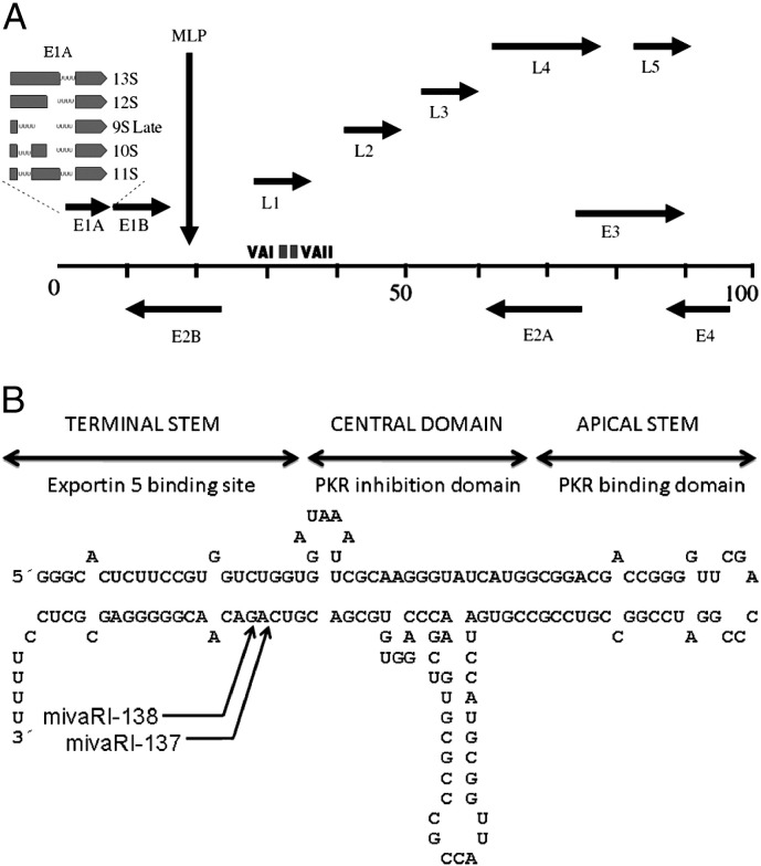
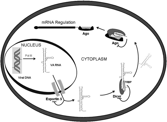
Adenovirus infection has a tremendous impact on the cellular silencing machinery. Adenoviruses express high amounts of non-coding virus associated (VA) RNAs able to saturate key factors of the RNA interference (RNAi) processing pathway, such as Exportin 5 and Dicer. Furthermore, a proportion of VA RNAs is cleaved by Dicer into viral microRNAs (mivaRNAs) that can saturate Argonaute, an essential protein for miRNA function. Thus, processing and function of cellular miRNAs is blocked in adenoviral-infected cells. However, viral miRNAs actively target the expression of cellular genes involved in relevant functions such as cell proliferation, DNA repair or RNA regulation. Interestingly, the cellular silencing machinery is active at early times post-infection and can be used to control the adenovirus cell cycle. This is relevant for therapeutic purposes against adenoviral infections or when recombinant adenoviruses are used as vectors for gene therapy. Manipulation of the viral genome allows the use of adenoviral vectors to express therapeutic miRNAs or to be silenced by the RNAi machinery leading to safer vectors with a specific tropism. This article is part of a "Special Issue entitled:MicroRNAs in viral gene regulation".
Copyright © 2011 Elsevier B.V. All rights reserved.
Figures
References
-
- Fabian M.R., Sonenberg N., Filipowicz W. Regulation of mRNA translation and stability by microRNAs. Annu. Rev. Biochem. 2010;79:351–379. - PubMed
-
- Bartel D.P. MicroRNAs: genomics, biogenesis, mechanism, and function. Cell. 2004;116:281–297. - PubMed
-
- Tomari Y., Zamore P.D. Perspective: machines for RNAi. Genes Dev. 2005;19:517–529. - PubMed
-
- Kim V.N. MicroRNA biogenesis: coordinated cropping and dicing. Nat. Rev. Mol. Cell Biol. 2005;6:376–385. - PubMed
-
- Kim V.N., Han J., Siomi M.C. Biogenesis of small RNAs in animals. Nat. Rev. Mol. Cell Biol. 2009;10:126–139. - PubMed
Publication types
MeSH terms
Substances
LinkOut - more resources
Full Text Sources
