. 2011 Sep;49(3):485-92.
doi: 10.1016/j.bone.2011.05.010. Epub 2011 May 18.

Spatiotemporal delivery of bone morphogenetic protein enhances functional repair of segmental bone defects

Affiliations

Spatiotemporal delivery of bone morphogenetic protein enhances functional repair of segmental bone defects

Yash M Kolambkar et al. Bone. 2011 Sep.

Abstract

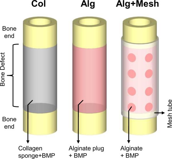
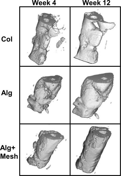
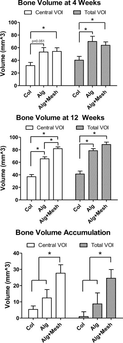
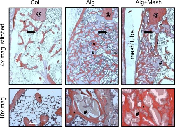
Osteogenic growth factors that promote endogenous repair mechanisms hold considerable potential for repairing challenging bone defects. The local delivery of one such growth factor, bone morphogenetic protein (BMP), has been successfully translated to clinical practice for spinal fusion and bone fractures. However, improvements are needed in the spatial and temporal control of BMP delivery to avoid the currently used supraphysiologic doses and the concomitant adverse effects. We have recently introduced a hybrid protein delivery system comprised of two parts: a perforated nanofibrous mesh that spatially confines the defect region and a functionalized alginate hydrogel that provides temporal growth factor release kinetics. Using this unique spatiotemporal delivery system, we previously demonstrated BMP-mediated functional restoration of challenging 8mm femoral defects in a rat model. In this study, we compared the efficacy of the hybrid system in repairing segmental bone defects to that of the current clinical standard, collagen sponge, at the same dose of recombinant human BMP-2. In addition, we investigated the specific role of the nanofibrous mesh tube on bone regeneration. Our results indicate that the hybrid delivery system significantly increased bone regeneration and improved biomechanical function compared to collagen sponge delivery. Furthermore, we observed that presence of the nanofiber mesh tube was essential to promote maximal mineralized matrix synthesis, prevent extra-anatomical mineralization, and guide an integrated pattern of bone formation. Together, these results suggest that spatiotemporal strategies for osteogenic protein delivery may enhance clinical outcomes by improving localized protein retention.


Figures

Figure 1
Figure 1
Schematic representations of the three protein delivery strategies used in this study. The first group consisted of a collagen sponge soaked with rhMBP-2 and implanted in the defect (Col). The second group consisted of an alginate hydrogel plug with rhBMP-2 (Alg). In the third group, alginate hydrogel containing rhBMP-2 was injected inside a perforated nanofiber mesh tube placed at the defect (Alg+Mesh).
Figure 2
Figure 2
Representative radiographs at 2, 4 and 12 weeks post-surgery. At 2 weeks, the Col specimens appeared to possess more bony tissue in the defect than the Alg and Alg+Mesh groups. However, at 4 and 12 weeks, defects in the two alginate groups demonstrated qualitatively higher bone formation. All defects in the three groups were bridged with bony tissue by 12 weeks. The newly formed bone in the alginate groups appeared more densely packed that that in the Col group. Note that the new bone formation was better contained within the defect in the Alg+Mesh group where the nanofiber mesh tube was present, compared to the Alg group. n = 9–10 defects per group.
Figure 3
Figure 3
Three-dimensional in vivo μCT images at 4 and 12 weeks. There was evidence of ample bone formation in the Col group specimens. Substantial bone formation in the Alg group occurred outside the defect. Alg+Mesh specimens, containing a nanofiber mesh tube, demonstrated continuous cylindrical bone distribution within the defect. n = 9–10 defects per group.
Figure 4
Figure 4
Quantitative μCT analysis of new bone volume. The Alg and Alg+Mesh groups had significantly higher bone formation than the Col group at both time points in the total VOI. At 12 weeks, the Alg+Mesh group possessed more bone in the central VOI region than the Alg group, consistent with the three-dimensional images. The Alg+Mesh group also saw the largest increase in the bone volume between 4 and 12 weeks. * indicates significantly different (p<0.05). n = 9–10 defects per group. Results are presented as mean±SEM.
Figure 5
Figure 5
Mean density (A) and connectivity density (B) of newly formed bone, obtained from μCT analysis. Mean density was significantly lower in the Alg+Mesh group compared to the other two groups, at both time points and in both VOIs. Connectivity density was the highest in the Alg+Mesh specimens at week 4, but no differences were observed at week 12. * indicates significantly different (p<0.05). n = 9–10 defects per group. Results are presented as mean±SEM.
Figure 6
Figure 6
Biomechanical properties of regenerated femurs obtained at 12 weeks by torsional testing. The Alg+Mesh specimens demonstrated higher maximum torque, torsional stiffness and work to failure than those in the Col group. No significant differences were observed in other comparisons. * indicates significantly different (p<0.05). n = 7–8 defects per group. Results are presented as mean±SEM.
Figure 7
Figure 7
Histological analysis at 12 weeks (4× and 10× magnification). H&E staining revealed improved bone regeneration in the two alginate groups, despite presence of residual alginate. The mesh tube in the Alg+Mesh group was able to constrain bone formation within the defect region. The black arrows are at located at the edges of the defects and point towards the center of the defects. The higher magnification images reveal osteocytes embedded in the newly formed bone (located with white arrows). Scale bars are 200 μm. @ - native bone end; * - regenerated bone; # - residual alginate. n = 2 defects per group.

References

    1. Cypher TJ, Grossman JP. Biological principles of bone graft healing. J Foot Ankle Surg. 1996;35:413–7. - PubMed
    1. Arrington ED, Smith WJ, Chambers HG, Bucknell AL, Davino NA. Complications of iliac crest bone graft harvesting. Clin Orthop Relat Res. 1996;329:300–9. - PubMed
    1. Berrey BH, Jr., Lord CF, Gebhardt MC, Mankin HJ. Fractures of allografts. Frequency, treatment, and end-results. J Bone Joint Surg Am. 1990;72:825–33. - PubMed
    1. Wheeler DL, Enneking WF. Allograft bone decreases in strength in vivo over time. Clin Orthop Relat Res. 2005;435:36–42. - PubMed
    1. Giannoudis PV, Dinopoulos H, Tsiridis E. Bone substitutes: an update. Injury. 2005;36(Suppl 3):S20–7. - PubMed

Publication types

MeSH terms

Substances