

Novel assessment of renal motion in children as measured via four-dimensional computed tomography
- PMID: 21621338
- PMCID: PMC3534728
- DOI: 10.1016/j.ijrobp.2011.03.046
Novel assessment of renal motion in children as measured via four-dimensional computed tomography
Abstract
Objectives: Abdominal intensity-modulated radiation therapy and proton therapy require quantification of target and organ motion to optimize localization and treatment. Although addressed in adults, there is no available literature on this issue in pediatric patients. We assessed physiologic renal motion in pediatric patients.
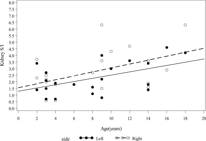
Methods and materials: Twenty free-breathing pediatric patients at a median age of 8 years (range, 2-18 years) with intra-abdominal tumors underwent computed tomography simulation and four-dimensional computed tomography acquisition (slice thickness, 3 mm). Kidneys and diaphragms were contoured during eight phases of respiration to estimate center-of-mass motion. We quantified center of kidney mass mobility vectors in three dimensions: anteroposterior (AP), mediolateral (ML), and superoinferior (SI).
Results: Kidney motion decreases linearly with decreasing age and height. The 95% confidence interval for the averaged minima and maxima of renal motion in children younger than 9 years was 5-9 mm in the ML direction, 4-11 mm in the AP direction, and 12-25 mm in the SI dimension for both kidneys. In children older than 9 years, the same confidence interval reveals a widening range of motion that was 5-16 mm in the ML direction, 6-17 mm in the AP direction, and 21-52 mm in the SI direction. Although not statistically significant, renal motion correlated with diaphragm motion in older patients. The correlation between diaphragm motion and body mass index was borderline (r = 0.52, p = 0.0816) in younger patients.
Conclusions: Renal motion is age and height dependent. Measuring diaphragmatic motion alone does not reliably quantify pediatric renal motion. Renal motion in young children ranges from 5 to 25 mm in orientation-specific directions. The vectors of motion range from 5 to 52 mm in older children. These preliminary data represent novel analyses of pediatric intra-abdominal organ motion.
Copyright © 2012 Elsevier Inc. All rights reserved.
Figures


References
-
- Dawson LA, Kavanagh BD, Paulino AC, et al. Radiation-Associated Kidney Injury. International Journal of Radiation Oncology*Biology*Physics. 2010;76:S108–S115. - PubMed
-
- Bolling T, Willich N, Ernst I. Late effects of abdominal irradiation in children: a review of the literature. Anticancer Res. 2010;30:227–231. - PubMed
-
- Moore JE, Jr., Ku DN. Pulsatile velocity measurements in a model of the human abdominal aorta under simulated exercise and postprandial conditions. J Biomech Eng. 1994;116:107–111. - PubMed
-
- Taremi M, Ringash J, Dawson LA. Upper abdominal malignancies: intensity-modulated radiation therapy. Front Radiat Ther Oncol. 2007;40:272–288. - PubMed
-
- Murtz P, Pauleit D, Traber F, et al. [Pulse triggering for improved diffusion-weighted MR imaging of the abdomen]. Rofo. 2000;172:587–590. - PubMed
Publication types
MeSH terms
Grants and funding
LinkOut - more resources
Full Text Sources
