. 2011 Jun 1;3(4):1425-42.
doi: 10.2741/e344.

Intercalated disc-associated protein, mXin-alpha, influences surface expression of ITO currents in ventricular myocytes

Affiliations

Intercalated disc-associated protein, mXin-alpha, influences surface expression of ITO currents in ventricular myocytes

Fu-Chi Chan et al. Front Biosci (Elite Ed). .

Abstract

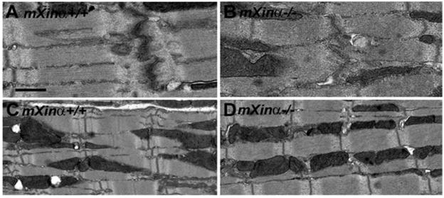
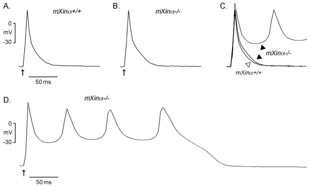
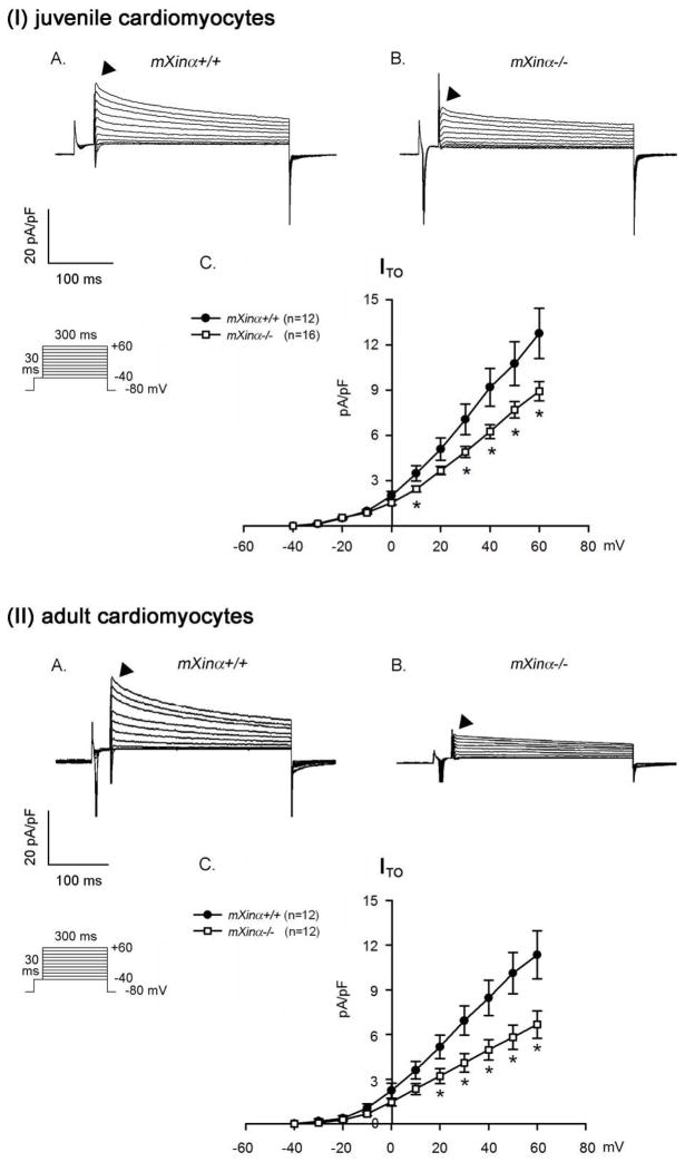
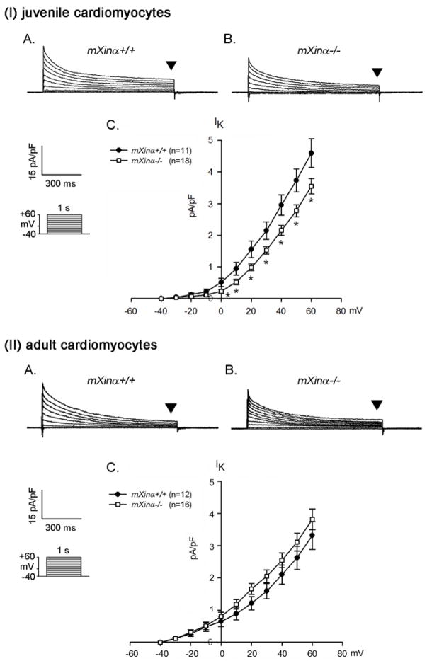
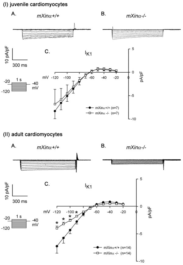
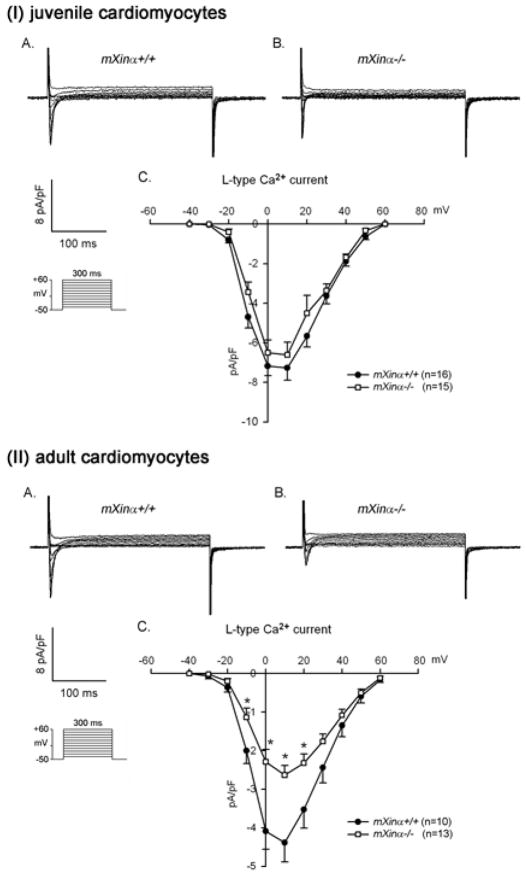
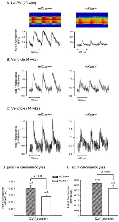
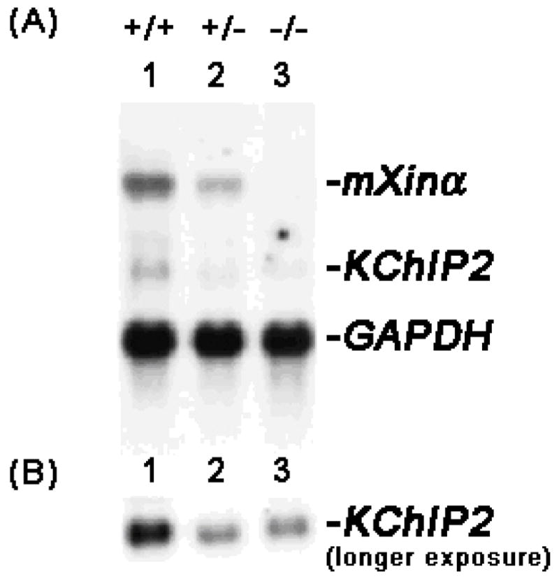
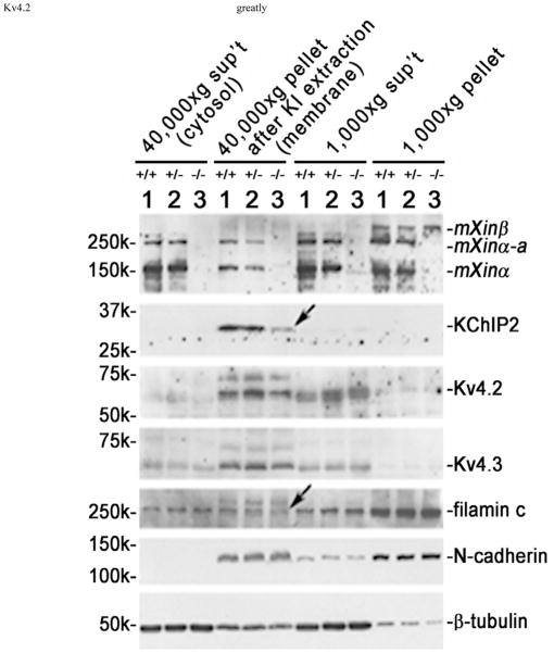
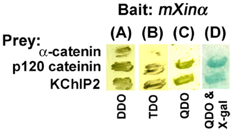
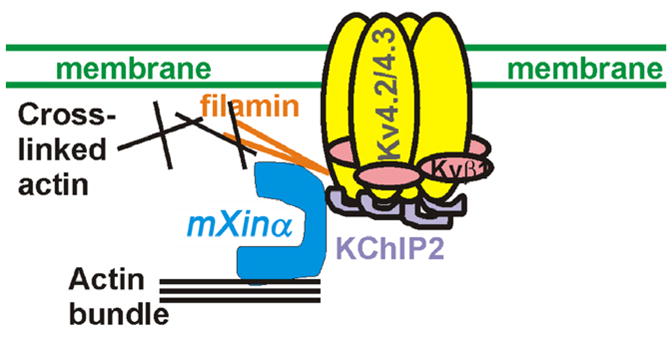
Mouse Xin-alpha (mXin-alpha) encodes a Xin repeat-containing, actin-binding protein localized to the intercalated disc (ICD). Ablation of mXin-alpha progressively leads to disrupted ICD structure, cardiac hypertrophy and cardiomyopathy with conduction defects during adulthood. Such conduction defects could be due to ICD structural defects and/or cell electrophysiological property changes. Here, we showed that despite the normal ICD structure, juvenile mXina-null cardiomyocytes (from 3~4-week-old mice) exhibited a significant reduction in the transient outward K+ current (ITO), similar to adult mutant cells. Juvenile but not adult mutant cardiomyocytes also had a significant reduction in the delayed rectifier K+ current. In contrast, the mutant adult ventricular myocytes had a significant reduction in the inward rectifier K+ current (IK1) on hyperpolarization. These together could account for the prolongation of action potential duration (APD) and the ease of developing early afterdepolarization observed in juvenile mXin-alpha-null cells. Interestingly, juvenile mXin-alpha-null cardiomyocytes had a notable decrease in the amplitude of intracellular Ca2+ transient and no change in the L-type Ca2+ current, suggesting that the prolonged APD did not promote an increase in intracellular Ca2+ for cardiac hypertrophy. Juvenile mXin-alpha-null ventricles had reduced levels of membrane-associated Kv channel interacting protein 2, an auxiliary subunit of ITO, and filamin, an actin cross-linking protein. We further showed that mXin-alpha interacted with both proteins, providing a novel mechanism for ITO surface expression.


Figures

Figure 1
Figure 1
Transmission electron microscopy. Comparisons of wild-type (A, C) and mXinα-null mouse hearts (B, D) at 1 month of age demonstrate no significant difference in the intercalated disc (A, B) and sarcomere (C, D) organizations, except less electron-density detected in mXinα-null ICD. Bar = 0.5 μm for A and B; 1 μm for C and D.
Figure 2
Figure 2
Action potential configurations of the typical juvenile wild-type (mXinα+/+) and mXinα−/− mouse ventricular myocytes. Panels A and B illustrate typical tracings of action potentials in mXinα+/+ and mXinα−/− ventricular myocytes, respectively, driven electrically at 1 Hz. Panel C shows the superimposed action potentials of panels A and B. Panel D illustrates another mXinα−/− ventricular myocyte developing a series of early afterdepolarizations (EAD) after a driven action potential. Upward arrows underneath the first driven action potential in each panel indicate time of electrical stimulation.
Figure 3
Figure 3
The ITO current densities of both juvenile and adult mXinα-null ventricular myocytes are significantly depressed as compared to those of wild-type counterparts. Membrane currents were elicited on depolarization from a holding potential of −80 mV to a test potential from −40 to +60 mV. Examples of transient outward currents (ITO) recorded in the (I) juvenile and (II) adult mXinα+/+ (Panel A) and mXinα−/− (Panel B) ventricular myocytes. Inset, various clamp protocols. Panel C summarizes current density-voltage (mean±SEM) relationships of ITO. n, Number of ventricular myocytes tested. * p<0.05, significant difference between wild-type and mXinα-null ventricular myocytes.
Figure 4
Figure 4
The delayed rectifier outward K+ current densities of juvenile but not adult mXinα-null ventricular myocytes are significantly depressed as compared to those of wild-type counterparts. Membrane currents were elicited on depolarization from a holding potential of −40 mV to a test potential from −40 to +60 mV. Examples of delayed rectifier K+ currents (indicated by downward solid triangles, IK) recorded in the (I) juvenile and (II) adult mXinα+/+ (Panel A) and mXinα−/− (Panel B) ventricular myocytes. Inset, various clamp protocols. Panel C summarizes current density-voltage (mean±SEM) relationships of IK. n, Number of ventricular myocytes tested. * p<0.05, significant difference between juvenile wild-type and mXinα-null ventricular myocytes.
Figure 5
Figure 5
Ba2+-sensitive inward rectifier K+ current (IK1) in juvenile and adult ventricular myocytes. Top panel (I) illustrates examples of a series of IK1 currents elicited on hyperpolarization in a wild-type (mXinα+/+) (A) and a mXinα-null myocyte (B). The clamp protocol is shown below the calibration bar. The differences in current densities before and after Ba2+ (mean ± SEM) were then plotted against the test potentials to obtain current-voltage relationship curves. No change in the IK1 was detected in juvenile mXinα-null ventricular myocytes (I) as compared to juvenile wild-type myocytes (C). In contrast, the IK1 currents of adult mXinα-null ventricular myocytes (II) were significantly depressed at test potentials more negative than −90 mV as compared to the wild-type counterparts (II). * p<0.05, significant difference between wild-type and mXinα-null ventricular myocytes.
Figure 6
Figure 6
The ICa,L current densities of juvenile mXinα-null ventricular myocytes are not significantly different from that of wild type counterparts, whereas the ICa,L current densities of adult mXinα-null ventricular myocytes are significantly reduced. Membrane currents were elicited on depolarization from a holding potential of −50 mV to a test potential from −40 to +60 mV. Examples of L-type Ca2+ current (ICa,L) recorded in the (I) juvenile and (II) adult mXinα+/+ (Panel A) and mXinα−/− (Panel B) ventricular myocytes. Inset, various clamp protocols. Panel C summarizes current density-voltage (mean±SEM) relationships of ICa,L. n, Number of ventricular myocytes tested. * p<0.05, significant difference between wild-type and mXinα-null ventricular myocytes. Note the current density-voltage curves of ICa,L originated from both mutant and wild type myocytes are peaked near +10 mV.
Figure 7
Figure 7
Intracellular Ca2+ concentration was decreased in both juvenile and adult mXinα-null cardiomyocytes. Panel A illustrates typical examples of Ca2+ transient in a wild type and a mXinα-null LA-PV cardiomyocytes (20-wks-old) as detected by Fluo-3 fluorescence (F/F0). Panels B and C illustrate examples of Ca2+ transient in juvenile (4 wks-old) and adult (14-wks-old) ventricular myocytes of wild type and mXinα-null mice, as detected by the Indo-1 fluorescence ratio (R410/485). Panels D and E summarize the average amplitudes of Ca2+ transients (R410/485) in juvenile (n = 7~9) and adult ventricular myocytes (n = 8~13). *p<0.05, significant difference in Ca2+ transient between wild-type and mXinα-null cells.
Figure 8
Figure 8
Down-regulation of KChIP2 in mXinα-deficient mouse hearts. (A) Total RNAs (10 μg each lane) prepared from wild-type (+/+, lane 1), heterozygous (+/−, lane 2) and homozygous (−/−, lane 3) mXinα mouse hearts were blotted and hybridized with a mixture of 32P-labeled mXinα cDNA and KChIP2 cDNA probes. For the RNA loading control, the same blot was further hybridized with GAPDH cDNA probe. (B) A longer exposure of the same Northern blot was shown to reveal the KChIP2 message band in mXinα-deficient hearts.
Figure 9
Figure 9
Juvenile mXinα-null ventricles exhibited significant decreases in both membrane-associated KChIP2 and filamin proteins (indicated by arrows). Total ventricular extracts were prepared as described under Materials and Methods in a low salt buffer, and spinned at 1,000×g centrifugation. The 1,000×g pellet contained nuclei, debris and low salt-resistant protein aggregates. The membrane vesicles, myofibrils and organelles in the 1,000×g supernatant (sup’t) were collected by 40,000×g centrifugation. After extracted with 6 M KI to remove myofibrils, the 40,000×g pellet and sup’t were termed “membrane” and “cytosolic” fractions, respectively. Western blot analysis was performed on these fractions prepared from ventricles of wild type (+/+, lane 1), heterozygous (+/−, lane 2) and homozygous (−/−, lane 3) littermates at 1 month of age. An equivalent volume from each fraction was loaded onto the SDS-PAGE gel, except 10 times concentrated sample of membrane fraction was used, and analyzed by Western blot with antibodies including U1013, anti-KChIP2, anti-Kv4.2, anti-Kv4.3, anti-filamin, anti-N-cadherin and anti-β-tubulin. The majority of Kv4.2 and Kv4.3, α-subunits, of ITO channel together with most of N-cadherin were detected in the membrane fraction, whereas most of β-tubulin was found in the cytosolic fraction.
Figure 10
Figure 10
The yeast two hybrid assays showed the interaction of mXinα with KChIP2. The α-catenin and p120 catenin preys were used in the same assay condition as a negative and a positive control, respectively. Yeast cells after transformation with both bait and prey plasmids were grown on double dropout (DDO) plate. Only the interaction occurring between bait and prey would support the cell growth on triple (TDO) and quadruple (QDO) dropout plates as well as the expression of β-galactosidase (blue color) on QDO & X-gal plate.
Figure 11
Figure 11
A schematic model suggesting that mXinα participates the assembly of ITO channel to the ICD of ventricular myocytes. In mice, the ITO channels reflect the assembly of Kv4.2/Kv4.3 α-subunits, Kvβ1 and KChIP2. A population of the ITO channels is known to localize at the ICD. Furthermore, it has been shown that the Kv4 pore-forming α-subunit is able to bind to filamin, and KChIP2. The associations with both proteins greatly enhance the surface expression of Kv4.2. In this study and our previous studies, we have clearly demonstrated that mXinα is capable of interacting with both KChIP2 and filamin. Through these interactions, mXinα likely influences surface expression of ITO channel.

Similar articles

Cited by

References

    1. Wang DZ, Reiter RS, Lin JL, Wang Q, Williams HS, Krob SL, Schultheiss TM, Evans S, Lin JJ. Requirement of a novel gene, Xin, in cardiac morphogenesis. Development. 1999;126(6):1281–94. - PubMed
    1. Lin JJ-C, Gustafson-Wagner EA, Sinn HW, Choi S, Jaacks SM, Wang DZ, Evans S, Lin JL-C. Structure, expression, and function of a novel intercalated disc protein, Xin. J Med Sci. 2005;25(5):215–222. - PMC - PubMed
    1. Huang HT, Brand OM, Mathew M, Ignatiou C, Ewen EP, McCalmon SA, Naya FJ. Myomaxin is a novel transcriptional target of MEF2A that encodes a Xin-related alpha-actinin-interacting protein. J Biol Chem. 2006;281(51):39370–9. - PubMed
    1. Sinn HW, Balsamo J, Lilien J, Lin JJ. Localization of the novel Xin protein to the adherens junction complex in cardiac and skeletal muscle during development. Dev Dyn. 2002;225(1):1–13. - PubMed
    1. Gustafson-Wagner EA, Sinn HW, Chen YL, Wang DZ, Reiter RS, Lin JL, Yang B, Williamson RA, Chen J, Lin CI, Lin JJ. Loss of mXinα, an intercalated disk protein, results in cardiac hypertrophy and cardiomyopathy with conduction defects. Am J Physiol Heart Circ Physiol. 2007;293(5):H2680–92. - PMC - PubMed

Publication types

LinkOut - more resources