Automated quantification of high-resolution CT scan findings in individuals at risk for pulmonary fibrosis
- PMID: 21622544
- PMCID: PMC3231958
- DOI: 10.1378/chest.10-2545
Automated quantification of high-resolution CT scan findings in individuals at risk for pulmonary fibrosis
Abstract
Background: Automated methods to quantify interstitial lung disease (ILD) on high-resolution CT (HRCT) scans in people at risk for pulmonary fibrosis have not been developed and validated.
Methods: Cohorts with familial pulmonary fibrosis (n = 126) or rheumatoid arthritis with and without ILD (n = 86) were used to develop and validate a computer program capable of quantifying ILD on HRCT scans, which imaged the lungs semicontinuously from the apices to the lung bases during end-inspiration in the prone position. This method uses segmentation, texture analysis, training, classification, and grading to score ILD.
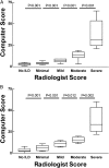
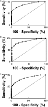
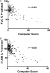
Results: Quantification of HRCT scan findings of ILD using an automated computer program correlated with radiologist readings and detected disease of varying severity in a derivation cohort with familial pulmonary fibrosis or their first-degree relatives. This algorithm was validated in an independent cohort of subjects with rheumatoid arthritis with and without ILD. Automated classification of HRCT scans as normal or ILD was significant in the derivation and validation cohorts (P < .001 and P < .001, respectively). Areas under receiver operating characteristic curves performed independently for each group were 0.888 for the derivation cohort and 0.885 for the validation cohort. Pulmonary function test results, including FVC and diffusion capacity, correlated with computer-generated HRCT scan scores for ILD (r = -0.483 and r = -0.532, respectively).
Conclusions: Automated computer scoring of HRCT scans can objectively identify ILD and potentially quantify radiographic severity of lung disease in populations at risk for pulmonary fibrosis.
Figures


References
-
- American Thoracic Society European Respiratory Society American Thoracic Society/European Respiratory Society International Multidisciplinary Consensus Classification of the Idiopathic Interstitial Pneumonias. This joint statement of the American Thoracic Society (ATS) and the European Respiratory Society (ERS) was adopted by the ATS board of directors, June 2001 and by the ERS Executive Committee, June 2001. Am J Respir Crit Care Med. 2002;165(2):277–304. - PubMed
-
- Lynch JP., III Computed tomographic scanning in sarcoidosis. Semin Respir Crit Care Med. 2003;24(4):393–418. - PubMed
-
- Chu SC, Horiba K, Usuki J, et al. Comprehensive evaluation of 35 patients with lymphangioleiomyomatosis. Chest. 1999;115(4):1041–1052. - PubMed
-
- Gochuico BR, Avila NA, Chow CK, et al. Progressive preclinical interstitial lung disease in rheumatoid arthritis. Arch Intern Med. 2008;168(2):159–166. - PubMed
-
- Warrick JH, Bhalla M, Schabel SI, Silver RM. High resolution computed tomography in early scleroderma lung disease. J Rheumatol. 1991;18(10):1520–1528. - PubMed
Publication types
MeSH terms
Grants and funding
LinkOut - more resources
Full Text Sources
Other Literature Sources
Medical

