. 2011 Jul 22;286(29):25710-8.
doi: 10.1074/jbc.M111.229179. Epub 2011 May 27.

Retroviral integrases promote fraying of viral DNA ends

Affiliations

Retroviral integrases promote fraying of viral DNA ends

Richard A Katz et al. J Biol Chem. .

Abstract

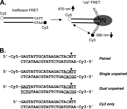
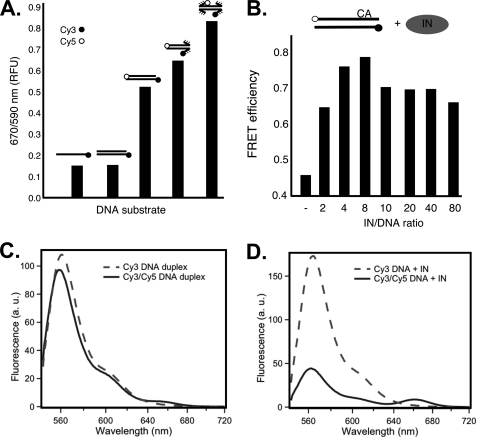
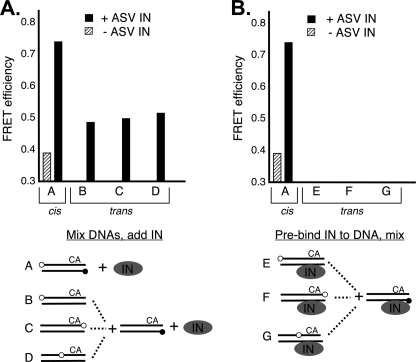
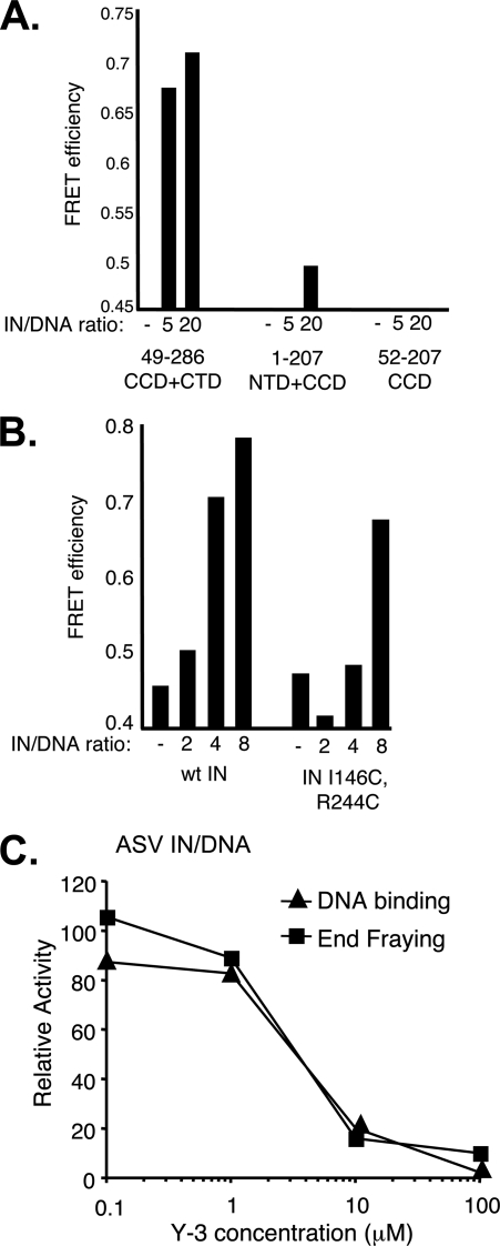
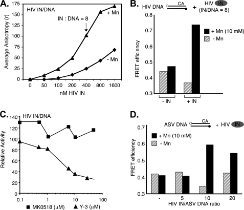
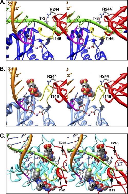
In the initial step of integration, retroviral integrase (IN) introduces precise nicks in the degenerate, short inverted repeats at the ends of linear viral DNA. The scissile phosphodiester bond is located immediately 3' of a highly conserved CA/GT dinucleotide, usually 2 bp from the ends. These nicks create new recessed 3'-OH viral DNA ends that are required for joining to host cell DNA. Previous studies have indicated that unpairing, "fraying," of the viral DNA ends by IN contributes to end recognition or catalysis. Here, we report that end fraying can be detected independently of catalysis with both avian sarcoma virus (ASV) and human immunodeficiency virus type 1 (HIV-1) IN proteins by use of fluorescence resonance energy transfer (FRET). The results were indicative of an IN-induced intramolecular conformational change in the viral DNA ends (cis FRET). Fraying activity is tightly coupled to the DNA binding capabilities of these enzymes, as follows: an inhibitor effective against both IN proteins was shown to block ASV IN DNA binding and end fraying, with similar dose responses; ASV IN substitutions that reduced DNA binding also reduced end fraying activity; and HIV-1 IN DNA binding and end fraying were both undetectable in the absence of a metal cofactor. Consistent with our previous results, end fraying is sequence-independent, suggesting that the DNA terminus per se is a major structural determinant for recognition. We conclude that frayed ends represent a functional intermediate in which DNA termini can be sampled for suitability for endonucleolytic processing.


Figures

FIGURE 1.
FIGURE 1.
DNA end fraying and experimental design. A, illustration of ASV viral DNA end fraying of terminal ∼4 bp by ASV IN as determined previously (4) and the predicted change in intramolecular (cis) FRET between donor (cyanine 3 (Cy3), filled circles) and acceptor (cyanine 5 (Cy5), open circles) fluorophores attached to the 5′ ends of a 22-bp oligodesoxyribonucleotide substrate. IN multimers, minimally dimers, are represented by a gray oval. B, sequences of the ASV-viral substrate duplexes used in these studies. Mismatched nucleotides at the termini are underlined. The small vertical arrows mark the normal sites of 3′ end processing by IN, in what is denoted the cleaved strand.
FIGURE 2.
FIGURE 2.
FRET changes depend on disruption of base pairing in DNA termini and the ratio of ASV IN to substrate DNA. A, FRET measured in the absence of IN. The bars show relative FRET values obtained with single- or double-stranded ASV-DNA end duplex substrates that contain only a donor fluorophore and duplex oligonucleotides that contain both donor and acceptor fluorophores with paired, or singly or doubly unpaired ends. B, FRET increase of the paired ASV-DNA end duplex substrate, optimal in IN excess, peaking as the ratio approaches 4. Concentration of the paired ASV-DNA end oligonucleotide was kept constant at 50 nm. Sequence is shown in Fig. 1. C, fluorescence emission spectra of matching (50 nm) samples of ASV-DNA duplex labeled with donor only (Cy3, dashed line) or donor and acceptor (Cy3/Cy5, solid line). D, emission spectra of the same DNA samples as in C, but in the presence of 400 nm ASV IN. Instrumental parameters, including excitation wavelength (535 nm) and bandwidth (2 nm for excitation and 4 nm for emission), were identical in both C and D. Labeling conventions in A and B are as in Fig. 1, with CA above the top line in B representing the conserved dinucleotide in the cleaved strand.
FIGURE 3.
FIGURE 3.
Distinguishing cis and trans FRET. A, duplex substrates and conditions were designed to distinguish cis versus trans IN-induced FRET. One fixed duplex contained only the FRET donor at the 5′ end of the noncleaved strand, as in the standard duplex substrate. A second set of duplexes contained only the acceptor fluorophore at three different positions on the cleaved strand (substrates B, C, and D). These substrates were designed to detect orientation-specific trans FRET. Equal amounts of separately labeled duplexes were first mixed, and then IN was added and the mixture left on ice for 5 min before FRET readings were taken. B, measurement of trans FRET efficiencies between duplexes containing only the acceptor or donor fluorophore and incubated separately with IN on ice for 5 min before mixing. FRET readings were taken immediately thereafter. The total substrate DNA concentration was 50 nm, and the ASV IN to DNA ratio was 4 in all experiments. The experimental schemes are illustrated below each graph with labeling conventions as in Figs. 1 and 2.
FIGURE 4.
FIGURE 4.
ASV IN CCD + CTD two-domain fragment retains fraying activity, and substitutions that reduce DNA binding also reduce end fraying. A, FRET efficiency as a function of the ratio of ASV IN-derived fragments to the viral DNA duplex. B, comparison of FRET efficiencies in the presence of increasing ratios of wild-type (wt) ASV IN and a full-length derivative that contains cysteine substitutions in the CCD and CTD, which reduce DNA binding. C, inhibition of ASV IN fraying and DNA binding by the Y-3. DNA binding was measured using a fluorescence polarization assay (17) in reactions that contained 50 nm of a 28- + 30-nucleotide substrate representing the recessed, processed U3 end of ASV DNA labeled with 6-carboxyfluorescein at the 5′ end of the cleaved strand, and 200 nm ASV IN. Y-3 was obtained from the NCI Chemical Repository through the Drug Synthesis and Chemistry Branch. The FRET was measured at the same IN:DNA ratio of 4. Data are plotted relative to control reactions that contained no Y-3. The same FRET substrate, at a constant 50 nm, was used in all three panels.
FIGURE 5.
FIGURE 5.
Detection of end fraying by HIV-1 IN. A, DNA binding measured using a fluorescence polarization assay (17) in reactions that contained 50 nm of a 20- + 22-nucleotide substrate representing the processed U5 end of HIV DNA, labeled with 6-carboxyfluorescein at the 5′ end of the cleaved strand as described in Fig. 4. Triangles, data for reactions that contained 10 mm MnCl2. Diamonds, data for reactions that contained no metal cofactor. B, relative FRET activity with a fully paired 22-bp HIV-1-DNA substrate duplex in the absence or presence a metal cofactor and IN:DNA ratio of 8. C, relative FRET activity of HIV-1 IN on an HIV-DNA substrate in the presence of Y-3 or MK0518 (raltegravir). D, FRET efficiencies with a fully paired 22-bp ASV-substrate duplex and HIV-1 IN at increasing IN:DNA ratios, in the absence or presence of a metal cofactor. Experimental designs are illustrated above the graphs, with labeling conventions as in Figs. 1 and 2. In both cases DNA duplex concentrations were at 50 nm.
FIGURE 6.
FIGURE 6.
ASV IN-mediated FRET changes are consistent with viral DNA end fraying. A, effects of dye positions on FRET efficiencies. Positioning of the Cy5 dye internally, 9 bp from either end of the 22-bp ASV substrate DNA, resulted in the expected increased FRET in the absence of IN compared with the substrate with terminal dyes. In the presence of ASV IN, there was a further increase in FRET with all substrates. The substrates with the internal dye allowed assessment of FRET changes at both ends of the viral substrate. Notably, an increase in FRET was detected on the non-CA end. B, FRET efficiencies and distance calculations compared between the ASV-DNA end duplex containing terminal or internal dyes in the absence or presence of ASV IN. Diagrams below highlight interactions that could contribute to increased FRET in the presence of IN. Use of the substrate with the internal Cy5 dye allows measurements uniquely at the CA end of the substrate, and this substrate was used to measure distance changes in the presence of IN. Apparent distances between the fluorophores (R) were calculated from the FRET efficiencies, in the presence or absence of IN, and are indicated below. Labeling conventions are as in Figs. 1 and 2.
FIGURE 7.
FIGURE 7.
Model of IN bound to a viral DNA end in the absence or presence of the inhibitors. A, the model of ASV IN was prepared using crystal structure of PFV IN bound to viral DNA as a template (Protein Data Bank ID code 3L2Q) (31, 40) and is portrayed as a stereo pair. The cleaved strand is shown as an orange ribbon, with the conserved CA residues in magenta. The noncleaved strand is in green and is held by cooperation of the CTD (red ribbon) and the flexible loop (yellow ribbon) adjacent to the active site in the CCD (blue ribbon). For clarity, the linkers between the NTD, core, and CTD domains are not shown. Active site residues that bind the divalent metal cofactors (purple sphere) as well as the two residues proposed to participate in positioning the noncleaved strand are portrayed in ball and stick: Ile146 (yellow) in the flexible loop and Arg244 in the CTD. The PFV residue Asn348, which is analogous to ASV Arg244, has been shown to make base-specific contacts; the Arg244 rotamer shown is in a position to make similar contact with the thymine at the −3 position in the noncleaved viral DNA strand. B, model of ASV IN bound to inhibitor Y-3. Color code is the same as in A except that the core domain ribbon is now shown in light blue. The Y-3 inhibitor is shown in space-filling representation in its previously established binding site (32), which induces a change in the position of the flexible loop and Ile146 (yellow) that preclude proper binding of the noncleaved DNA strand. C, model of HIV-1 IN bound to both Y-3 and raltegravir. Colors are as above, except that the HIV-1 CCD is in cyan. To position Y-3, the HIV-1 intasome model with bound raltegravir (21) was superimposed onto the ASV IN catalytic core bound to Y-3. As with ASV IN, Y-3 is predicted to interfere sterically with binding of the noncleaved strand of HIV viral DNA. In contrast, the presence of viral DNA in the complex enhances raltegravir binding to HIV-1 IN, but such binding is predicted to interfere with target DNA binding. Importantly, Y-3 and raltegravir are predicted to bind on opposite sides of the flexible loop.

References

    1. Vandegraaff N., Engelman A. (2007) Expert Rev. Mol. Med. 9, 1–19 - PubMed
    1. Skalka A. M., Katz R. A. (2005) Cell Death Differ. 12, 971–978 - PubMed
    1. Yi J., Asante-Appiah E., Skalka A. M. (1999) Biochemistry 38, 8458–8468 - PubMed
    1. Katz R. A., DiCandeloro P., Kukolj G., Skalka A. M. (2001) J. Biol. Chem. 276, 34213–34220 - PubMed
    1. Oh J., Chang K. W., Hughes S. H. (2008) J. Virol. 82, 11480–11483 - PMC - PubMed

Publication types

LinkOut - more resources