. 2011 Jul 29;286(30):26507-15.
doi: 10.1074/jbc.M111.224634. Epub 2011 May 27.

Oxygen supply from the bird's eye perspective: globin E is a respiratory protein in the chicken retina

Affiliations

Oxygen supply from the bird's eye perspective: globin E is a respiratory protein in the chicken retina

Miriam Blank et al. J Biol Chem. .

Abstract

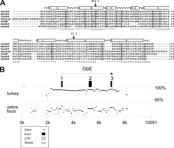
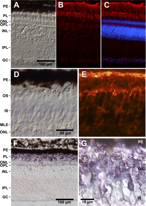
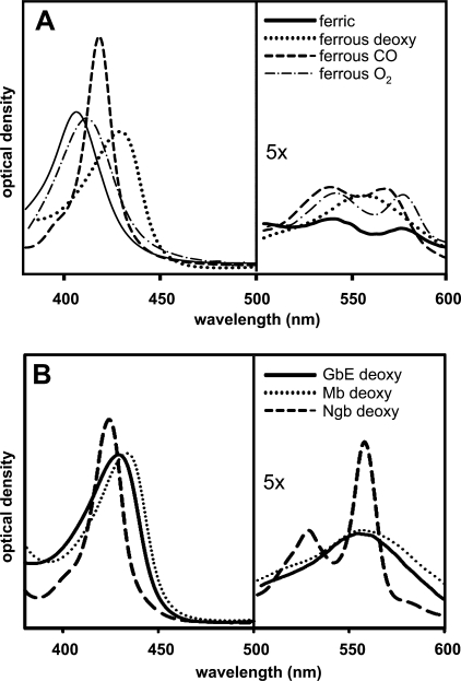
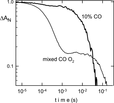
The visual process in the vertebrate eye requires high amounts of metabolic energy and thus oxygen. Oxygen supply of the avian retina is a challenging task because birds have large eyes, thick retinae, and high metabolic rates but neither deep retinal nor superficial capillaries. Respiratory proteins such as myoglobin may enhance oxygen supply to certain tissues, and thus the mammalian retina harbors high amounts of neuroglobin. Globin E (GbE) was recently identified as an eye-specific globin of chicken (Gallus gallus). Orthologous GbE genes were found in zebra finch and turkey genomes but appear to be absent in non-avian vertebrate classes. Analyses of globin phylogeny and gene synteny showed an ancient origin of GbE but did not help to assign it to any specific globin type. We show that the photoreceptor cells of the chicken retina have a high level of GbE protein, which accumulates to ∼10 μM in the total eye. Quantitative real-time RT-PCR revealed an ∼50,000-fold higher level of GbE mRNA in the eye than in the brain. Spectroscopic analysis and ligand binding kinetics of recombinant chicken GbE reveal a penta-coordinated globin with an oxygen affinity of P(50) = 5.8 torrs at 25 °C and 15 torrs at 41 °C. Together these data suggest that GbE helps to sustain oxygen supply to the avian retina.


Figures

FIGURE 1.
FIGURE 1.
Conservation of GbE protein and gene. A, comparison of eye globins of chicken (GgaGbE), turkey (MgaGbE) and zebra finch (TguGbE) with chicken Cygb (GgaCygb), myoglobin (GgaMb), hemoglobin α (GgaHbA), and β (GgaHbB). The sperm whale myoglobin structure is superimposed in the upper row, and residues conserved in all these globins are shaded. B, percent identity plot showing the comparison chicken GbE with turkey and zebra finch GbE genes and their ± 3 kb flanking regions. The G. gallus GbE was used as reference; in the upper row, the exons are boxed, with the black boxes representing the coding sequences. The transcriptional orientation is indicated by the arrow. Interspecies sequence identities are shown as horizontal bars on a 50–100% scale.
FIGURE 2.
FIGURE 2.
Expression of GbE protein and RNA. A, expression of GbE and Ngb mRNA in chicken brain and eye, as measured by quantitative real-time RT-PCR. Values are means of three independent replicates (n = 3), and the standard deviations are given. Amounts of globin mRNA are given as copy number per μg of total RNA. Note the logarithmic scale of the y axis. B, Western blot detection of GbE protein in chicken eye. On the left lane, 0.5 μg of purified recombinant GbE was applied; on the right lane, 100 μg of total proteins extracted from eye were loaded. Protein was detected employing a specific anti-GbE antibody. GbE content was estimated by ImageJ to be 1.6 ± 0.1 μg GbE/mg of total eye proteins (n = 3).
FIGURE 3.
FIGURE 3.
Localization of GbE protein and mRNA in the chicken retina. A–C, longitudinal cryosections of the chicken retina. PE, pigment epithelium; PL, layer of outer and inner segments of photoreceptor cells; ONL, outer nuclear layer; OPL, outer plexiform layer; INL, inner nuclear layer; IPL, inner plexiform layer; GC, layer of ganglion cells. A, bright field microscopy image. Scale bar, 100 μm. B, indirect anti-GbE immunofluorescence. Bright immunofluorescence is visible in the pigment epithelium and the outer segments of photoreceptors; weak staining is visible in the outer plexiform layer and the ganglion cells. C, merged figure showing staining of the nuclei with Hoechst dye 33258. D and E, higher magnification of the photosensitive outer segments (OS) shows anti-GbE immunofluorescence of the cytoplasm surrounding the stacks of membrane-enclosed disks (E). D, bright field image. Scale bar, 20 μm. IS, inner segments; MLE, membrana limitans externa. F and G, immunohistological staining employing a secondary antibody coupled with alkaline phosphatase shows strong staining of the outer segments.
FIGURE 4.
FIGURE 4.
Absorbance spectra of GbE. A, spectral forms of ferric (solid line), ferrous deoxygenated (dotted line), ferrous CO-form (dashed line), and ferrous oxygenated (dot-dash line) of recombinant GbE were read from 380 to 600 nm. The protein concentration was ∼0.15 mg/ml. B, comparison of spectra of deoxygenated GbE (solid line), Mb (dotted line), and Ngb (dashed line).
FIGURE 5.
FIGURE 5.
Flash photolysis kinetics of GbE. After photodissociation of CO, the CO recombination showed a slight heterogeneity (bold line) in the recombination kinetics. The use of a mixed CO/O2 atmosphere (thin line) allows a determination of the oxygen on and off rates. GbE has the characteristic of classical globins, where CO binds more tightly, but O2 binds more rapidly. Photolysis of CO produces the unliganded (penta-coordinated) transient; the rapid phase is essentially oxygen binding, and the slow phase is the replacement of oxygen by CO. Data are presented on a log-log scale as the normalized change in absorption versus time, for curves measured at 25 °C, pH 8.0.

References

    1. Dickerson A. E., Greis I. (1983) Hemoglobin: Structure, Function, Evolution, and Pathology, Benjamin-Cummings Publishing Company, Inc., Menlo Park, CA
    1. Wittenberg J. B., Wittenberg B. A. (2003) J. Exp. Biol. 206, 2011–2020 - PubMed
    1. Burmester T., Weich B., Reinhardt S., Hankeln T. (2000) Nature 407, 520–523 - PubMed
    1. Burmester T., Hankeln T. (2009) J. Exp. Biol. 212, 1423–1428 - PubMed
    1. Hankeln T., Ebner B., Fuchs C., Gerlach F., Haberkamp M., Laufs T. L., Roesner A., Schmidt M., Weich B., Wystub S., Saaler-Reinhardt S., Reuss S., Bolognesi M., De Sanctis D., Marden M. C., Kiger L., Moens L., Dewilde S., Nevo E., Avivi A., Weber R. E., Fago A., Burmester T. (2005) J. Inorg. Biochem. 99, 110–119 - PubMed

Publication types