. 2011 Jul 22;286(29):26050-60.
doi: 10.1074/jbc.M111.248765. Epub 2011 May 26.

Dimer interface of the effector domain of non-structural protein 1 from influenza A virus: an interface with multiple functions

Affiliations

Dimer interface of the effector domain of non-structural protein 1 from influenza A virus: an interface with multiple functions

James M Aramini et al. J Biol Chem. .

Abstract

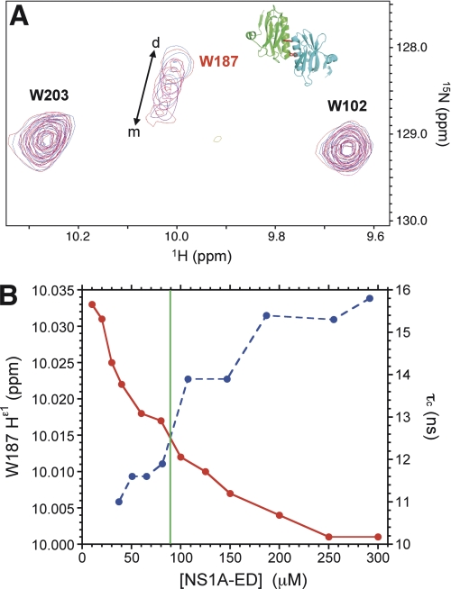
Non-structural protein 1 from influenza A virus, NS1A, is a key multifunctional virulence factor composed of two domains: an N-terminal double-stranded RNA (dsRNA)-binding domain and a C-terminal effector domain (ED). Isolated RNA-binding and effector domains of NS1A both exist as homodimers in solution. Despite recent crystal structures of isolated ED and full-length NS1A proteins from different influenza virus strains, controversy remains over the actual biologically relevant ED dimer interface. Here, we report the biophysical properties of the NS1A ED from H3N2 influenza A/Udorn/307/1972 (Ud) virus in solution. Several lines of evidence, including (15)N NMR relaxation, NMR chemical shift perturbations, static light scattering, and analytical sedimentation equilibrium, demonstrate that Ud NS1A ED forms a relatively weak dimer in solution (K(d) = 90 ± 2 μm), featuring a symmetric helix-helix dimer interface. Mutations within and near this interface completely abolish dimerization, whereas mutations consistent with other proposed ED dimer interfaces have no effect on dimer formation. In addition, the critical Trp-187 residue in this interface serves as a sensitive NMR spectroscopic marker for the concentration-dependent dimerization of NS1A ED in solution. Finally, dynamic light scattering and gel shift binding experiments demonstrate that the ED interface plays a role in both the oligomerization and the dsRNA binding properties of the full-length NS1A protein. In particular, mutation of the critical tryptophan in the ED interface substantially reduces the propensity of full-length NS1A from different strains to oligomerize and results in a reduction in dsRNA binding affinity for full-length NS1A.


Figures

FIGURE 1.
FIGURE 1.
Multiple sequence alignment of NS1A proteins from various influenza virus strains. Multiple sequence alignment of full-length NS1A from influenza A/Udorn/307/1972 (H3N2), influenza A/Puerto Rico/8/1934 (H1N1), influenza A/Duck/Alberta/60/1976 (H12N5), and influenza A/Vietnam/1203/2004 (H5N1) is shown. Conserved residues are shown in red. The RBD and ED domains are outlined in blue and green, respectively. Residue numbering and secondary structural elements from the structures of Ud NS1A RBD (PDB ID 1NS1) and Ud NS1A ED (PDB ID 3EE9) are shown above the sequence. The putative strand-strand and helix-helix ED dimer interfaces are boxed in yellow and blue, respectively. Triangles denote the residues mutated in this study, and the position of Trp-187, important for dimerization of NS1A ED, is marked by an asterisk.
FIGURE 2.
FIGURE 2.
Assessment of wild type and mutant Ud NS1A ED oligomerization state using 15N relaxation. A, plots of rotational correlation time (τc) determined from 15N T1 and T2 relaxation data as a function of protein molecular weight (MW). Blue, wild type Ud NS1A(85–215) and mutants (0.3–0.7 mm in pH 6.9 buffer, 300 K); red, known monomeric proteins solved in the NESG project (38). All NS1A ED samples were U-5%-13C,100%-15N-labeled except W187R-NC, U-13C,15N-labeled [W187R]Ud NS1A(85–215), and WT-ILVFY, [U-2H,13C,15N; 1H-Ile-δ1,Leu-δ,Val-γ,Phe,Tyr]Ud NS1A(85–215); additional isotopic enrichment produces an expected shift to longer τc values. B, effect of mutations on NS1A ED oligomerization state mapped onto literature structures of PR8 NS1A ED (left; PDB ID 2GX9 (20)) and Ud NS1A ED (right; PDB ID 3EE9 (22)). Side chains for mutated residues are colored as follows: red, mutants that are monomeric; yellow, mutants that result in either monomer or dimer formation; blue, mutants that are dimeric.
FIGURE 3.
FIGURE 3.
NMR chemical shift perturbations in Ud NS1A ED resulting from the mutation of Trp-187. A plot of combined 15N and 1H chemical shift perturbations as a function of residue between wild type Ud NS1A(85–215) and [W187R]Ud NS1A(85–215), obtained from 800 MHz 1H-15N TROSY-HSQC spectra of 0.3–0.4 mm U-5%-13C,100%-15N-labeled protein samples in pH 6.9 buffer at 300 K, is shown. Inset: residues with Δδcomp >0.1 ppm are mapped in red onto the dimer structure of Ud NS1A ED (PDB ID 3EE9 (22)) and cluster within or near the helix-helix dimer interface.
FIGURE 4.
FIGURE 4.
Analytical gel filtration and static light scattering data for wild type and [W187R]Ud NS1A ED. Data were collected at room temperature on [U-5%-13C,100%-15N]Ud NS1A(85–215) (black, 20 μm; red, 400 μm) and U-5%-13C,100%-15N-labeled [W187R]Ud NS1A(85–215) (blue, 20 μm; green, 400 μm) in pH 6.9 buffer. Note the shift to lower elution volume for the concentrated sample of wild type Ud NS1A ED, indicative of a higher molecular weight species.
FIGURE 5.
FIGURE 5.
Trp-187 is a spectroscopic marker for dimerization of Ud NS1A ED. A, overlay of 600 MHz 1H-15N HSQC spectra of Ud NS1A(85–215) at various protein concentrations (pH 6.9 buffer, 300 K) showing the tryptophan Hϵ1/Nϵ1 region. Inset: positions of Trp-187 in the dimer structure of Ud NS1A ED (PDB ID 3EE9 (22)). B, plots of Trp-187 Hϵ1 chemical shift (red) and τc (blue) as a function of Ud NS1A ED concentration. The green line indicates the Kd at ∼90 μm for dimer formation of Ud NS1A(85–215) determined by sedimentation equilibrium.
FIGURE 6.
FIGURE 6.
Solution NMR studies of [W187R]Ud and wild type Ud NS1A(85–215). A, ribbon diagrams of the lowest energy (CNS) conformer from the final solution NMR structure ensemble of [W187R]Ud NS1A(85–215) (PDB ID 2KKZ). The α-helices and β-strands are shown in cyan and magenta, respectively. The side chain of the mutant Arg-187 is shown in red. For clarity, the highly disordered residues at the C terminus of the molecule (after Gly-204) have been omitted. B, strips from the 800-MHz three-dimensional 13C-edited (left) and 15N-edited (right) NOESY spectra of 0.43 mm [U-2H,13C,15N; 1H-Ile-δ1,Leu-δ,Val-γ,Phe,Tyr]Ud NS1A(85–215) showing intermolecular NOESY contacts at the helix-helix dimer interface of Ud NS1A ED. C, close-up view of the helix-helix dimer interface from the x-ray crystal structure of Ud NS1A ED (PDB ID 3EE9 (22)) showing intermolecular Val-180-Trp-187 side chain contacts.
FIGURE 7.
FIGURE 7.
Effects of mutating tryptophan on the oligomerization of full-length NS1A. Plots of effective molecular weight obtained from dynamic light scattering versus NS1A concentration are shown. A, Ud NS1A(1–215). Blue, wild type protein; red, [W187A] mutant. B, VN NS1A(1–215). Blue, wild type protein; red, [W182A] mutant. For both influenza strains, mutation of the conserved tryptophan dramatically reduces the aggregation property of full-length NS1A.
FIGURE 8.
FIGURE 8.
EMSA gel shift dsRNA binding experiments on full-length and truncated Ud NS1A. A, wild type Ud NS1A(1–215). B, [W187R]Ud NS1A(1–215). C, Ud NS1A(1–73). The protein monomer concentrations (in nm) are indicated in each lane. The experiment was repeated twice, yielding identical results each time. Approximate monomer protein concentrations required for 50% complex formation with dsRNA are as follows: wild type, 14 nm; [W187R]Ud mutant, 28 nm; NS1A RBD, 65 nm.
FIGURE 9.
FIGURE 9.
Working model for the oligomerization of NS1A upon binding to dsRNA. Views into (top) and along (bottom) the dsRNA helical axis of a model of the complex between full-length NS1A and dsRNA (black) are shown. The RBD and ED domains are rendered as cylinders and surfaces, respectively. Domains corresponding to full-length NS1A dimers (mediated by RBD-RBD interactions) are shown in similar colors; ED subunits are labeled. The model was made using the coordinates from the x-ray crystal structures of PR8 NS1A RBD-dsRNA complex (PDB ID 2ZKO (19)), preserving the known dsRNA binding-topology observed in this complex, and Ud NS1A ED (PDB ID 3EE9 (22)), featuring a helix-helix dimer interface, by overlaying multiple copies of the RBD-dsRNA complex coordinates onto canonical dsRNA and manually docking the ED coordinates onto separate RBDs. In this schematic model, the protein wraps around the dsRNA via its RBD, forming a tube with the dsRNA in the center. Each RBD dimer binds the major groove of dsRNA (19) via highly conserved tracks of amino acid residues from helices 2 and 2′ (18). The oligomer is held together by intermolecular contacts mediated by the helix-helix interface between adjacent ED domains. Left inset: view of the dsRNA-binding epitope of NS1A RBD. The relative orientation of the dsRNA parallel to the axes of helices 2 and 2′ is determined by highly conserved tracks of basic residues (18) and other polar dsRNA-binding residues (19) lining the dsRNA-binding face, shown in dark and light blue, respectively. Right inset: view of the intermolecular ED-ED interface (22). The side chains of the critical Trp-187 residues from each subunit are shown in red and indicated by an arrow.

References

    1. Taubenberger J. K., Morens D. M. (2006) Emerg. Infect. Dis. 12, 15–22 - PMC - PubMed
    1. Krug R. M., Aramini J. M. (2009) Trends Pharmacol. Sci. 30, 269–277 - PMC - PubMed
    1. Das K., Aramini J. M., Ma L. C., Krug R. M., Arnold E. (2010) Nat. Struct. Mol. Biol. 17, 530–538 - PMC - PubMed
    1. Hale B. G., Randall R. E., Ortín J., Jackson D. (2008) J. Gen. Virol. 89, 2359–2376 - PubMed
    1. Min J. Y., Krug R. M. (2006) Proc. Natl. Acad. Sci. U.S.A. 103, 7100–7105 - PMC - PubMed

Publication types

Associated data