. 2011 Jul;77(14):4849-58.
doi: 10.1128/AEM.02599-10. Epub 2011 May 27.

Assembly of minicellulosomes on the surface of Bacillus subtilis

Affiliations

Assembly of minicellulosomes on the surface of Bacillus subtilis

Timothy D Anderson et al. Appl Environ Microbiol. 2011 Jul.

Retraction in

Abstract

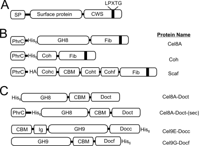
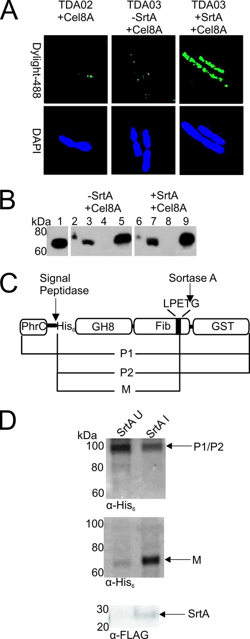
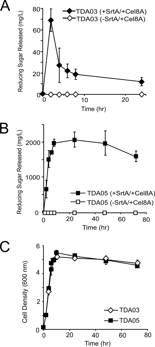
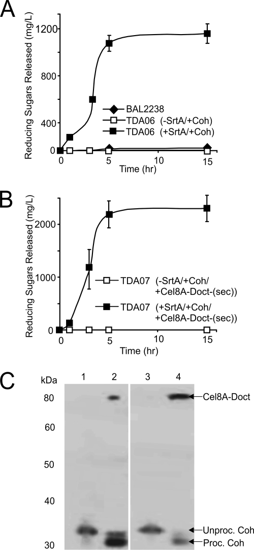
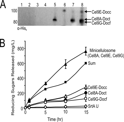
To cost-efficiently produce biofuels, new methods are needed to convert lignocellulosic biomass into fermentable sugars. One promising approach is to degrade biomass using cellulosomes, which are surface-displayed multicellulase-containing complexes present in cellulolytic Clostridium and Ruminococcus species. In this study we created cellulolytic strains of Bacillus subtilis that display one or more cellulase enzymes. Proteins containing the appropriate cell wall sorting signal are covalently anchored to the peptidoglycan by coexpressing them with the Bacillus anthracis sortase A (SrtA) transpeptidase. This approach was used to covalently attach the Cel8A endoglucanase from Clostridium thermocellum to the cell wall. In addition, a Cel8A-dockerin fusion protein was anchored on the surface of B. subtilis via noncovalent interactions with a cell wall-attached cohesin module. We also demonstrate that it is possible to assemble multienzyme complexes on the cell surface. A three-enzyme-containing minicellulosome was displayed on the cell surface; it consisted of a cell wall-attached scaffoldin protein noncovalently bound to three cellulase-dockerin fusion proteins that were produced in Escherichia coli. B. subtilis has a robust genetic system and is currently used in a wide range of industrial processes. Thus, grafting larger, more elaborate minicellulosomes onto the surface of B. subtilis may yield cellulolytic bacteria with increased potency that can be used to degrade biomass.


Figures

Fig. 1.
Fig. 1.
Schematic showing proteins used in this study. (A) A generalized substrate of the SrtA sortase that contains an N-terminal secretory peptide (SP) and C-terminal cell wall sorting signal (CWS) with the LPXTG sorting motif. (B) Specific surface proteins that were anchored to the cell wall by SrtA. Each protein contains the SP derived from B. subtilis PhrC, followed by a hexahistidine (His6) or human influenza hemagglutinin (HA) tag. At their C termini, each protein contains the CWS from the S. aureus fibronectin binding protein B (Fib). Cel8A is the endoglucanase A from C. thermocellum and contains the family-8 glycoside hydrolyase (GH) module. Coh is the type I cohesin from the C. thermocellum CipA protein. Scaf contains several domains and has been described previously (15). It contains the type I cohesin from C. cellulolyticum CipC (Cohc), a carbohydrate binding module (CBM), a type I cohesin from C. thermocellum CipA (Coht), and a type I cohesin from R. flavefaciens ScaB (Cohf). (C) Schematic of the dockerin-containing cellulase enzymes that were displayed on the surface of B. subtilis. Proteins purified from E. coli include the following: Cel9E-Docc, the C. cellulolyticum exoglucanase Cel9E fused to its native dockerin and containing a family-9 GH, an immunoglobulinlike module (IG), and CBM (15); Cel9G-Docf, C. cellulolyticum endoglucanase Cel9G that contains a family-9 GH and CBM, fused to a type I dockerin from R. flavefaciens ScaA (15); and Cel8A-Doct, the Cel8A endoglucanase from C. thermocellum fused to the CBM and dockerin modules derived from the C. thermocellum xylanase Xyn10B protein. CelA-Doct-(sec) was used to assemble the cohesin-cellulase complex through coexpression.
Fig. 2.
Fig. 2.
Cel8A is successfully displayed on the surface of B. subtilis. (A) Immunofluorescence micrographs of B. subtilis strain TDA03 displaying His6-tagged Cel8A. Left, cells of strain TDA02 expressing Cel8A. Middle, cells of strain TDA03 expressing only Cel8A. Right, cells of strain TDA03 expressing both SrtA and Cel8A. Cells were probed for the presence of Cel8A on the surface with mouse anti-His6 serum and fluorescently stained anti-mouse IgG conjugated to Dylight-488. 4′,6-diamidino-2-phenylindole (DAPI) was used to stain the DNA. In images containing larger numbers of cells, a similar display pattern was observed. (B) Immunoblot analysis of the cellular localization of Cel8A in strain TDA03. Lanes: 1, purified Cel8A; 2 and 6, lysed whole cells; 3 and 7, lysozyme-solubilized cell wall; 4 and 8, membrane/cytosol; 5 and 9, precipitated secreted protein. Samples were probed with a mouse anti-His6 antibody. Lanes 2 to 5 represent samples in which SrtA was not expressed. Lanes 6 to 9 represent samples in which SrtA was expressed. (C) Diagram showing the Cel8A-GST protein used to track processing by SrtA. The expected forms of the protein include P1, the unprocessed full-length precursor; P2, the precursor protein after cleavage by the signal peptidase; and M, the mature protein after cleavage of the CWS by SrtA. (D). Immunoblots of cell fractions of strain TDA08 expressing Cel8A-GST and/or SrtA. Top, SDS-released cytoplasmic fractions in cells in which SrtA expression has not been induced (SrtA U, left column) or has been induced (SrtA I, right column). Middle, blot of cell wall extracts that had been digested with mutanolysin. Bottom, detection of SrtA expression in the SDS-treated cytoplasmic and membrane fractions using an anti-FLAG antibody.
Fig. 3.
Fig. 3.
Eliminating the WprA protease increases the cellulolytic activity of B. subtilis cells displaying the Cel8A cellulase. (A) Cellulase activity of TDA03 cells during growth. Growth cultures of cells displaying Cel8A (+SrtA/+Cel8A) or not displaying CelA (-SrtA/+Cel8A) were periodically collected and washed and their abilities to degrade carboxymethyl cellulose determined by measuring the amount of reducing sugars that were released. (B) Identical to panel A, except that strain TDA05 was used, in which the WprA cell wall-associated protease has been genetically deleted. (C) Corresponding growth curves of TDA03 and TDA05 as a function of time. Activity profiles were performed in triplicate. The reported error is the standard deviation of these measurements.
Fig. 4.
Fig. 4.
Assembly of a cohesin:cellulase complex on the surface of B. subtilis either by adding purified cellulase or by coexpressing each component. (A) Display of the cohesin:cellulase complex after adding purified cellulase enzyme. Cultures of TDA06 induced to display Coh were grown for various amounts of time. Purified Cel8A-Doct was then added, and the ability of washed cells to degrade CMC was determined. (B) Display of the cohesin:cellulase complex by coexpressing its components. Strain TDA07 was induced to express SrtA, Coh, and Cel8A-Doct-(sec) and periodically collected during the expression, and the ability of the cells to degrade CMC was determined. Experiments in panels A and B were performed in triplicate, and the error reported is the standard deviation. (C) Immunoblot of cell wall fractions of strain TDA06 (lanes 1 and 2) exposed to purified Cel8A-Doct and strain TDA07 (lanes 3 and 4) expressing Coh and Cel8A-Doct-(sec). Lane 1, TDA06 -SrtA/+Coh/+Cel8A-Doct. Lane 2, TDA06 +SrtA/+Coh/+Cel8A-Doct. Lane 3, TDA07 -SrtA/+Coh:Cel8A-Doct-(sec). Lane 4, TDA07+SrtA/+Coh:Cel8A-Doct-(sec). Unproc., unprocessed; Proc., processed. Samples were probed using a mouse anti-His6 antibody.
Fig. 5.
Fig. 5.
Assembly of a surface-displayed minicellulosome that contains three enzymes. (A) Immunoblot analysis of the cell wall of cells of strain TDA09 expressing Scaf (lanes 1 to 4) only or both SrtA and Scaf (lanes 5 to 8). Cells were incubated individually with Cel8A-Doct (lanes 1 and 5), Cel9E-Docc (lanes 2 and 6), Cel9G-Dockf (lanes 3 and 7), or all three cellulases (lanes 4 and 8). The cell walls were then solubilized and the proteins probed with an anti-His6 antibody. (B) Whole-cell activity of cells displaying individual enzymes or a minicellulosome. Cultures of strain TDA09 expressing Scaf and/or SrtA were periodically collected and incubated with purified Cel8A-Doct, Cel9E-Docc, and/or Cel9G-Docf protein. After washing, activity against HCl-treated amorphous cellulose was determined. The curve labeled “Sum” is the sum of the enzymatic activities of cells incubated with only a single type of enzyme. Whole-cell cellulase assays were performed in triplicate, and the standard deviation of these measurements is used to represent the error.

References

    1. Bayer E. A., Morag E., Lamed R. 1994. The cellulosome—a treasure-trove for biotechnology. Trends Biotechnol. 12: 379–386 - PubMed
    1. Bayer E. A., Setter E., Lamed R. 1985. Organization and distribution of the cellulosome in Clostridium thermocellum. J. Bacteriol. 163: 552–559 - PMC - PubMed
    1. Bayer E. A., Shimon L. J., Shoham Y., Lamed R. 1998. Cellulosomes—structure and ultrastructure. J. Struct. Biol. 124: 221–234 - PubMed
    1. Bron S. 1990. Plasmids, p. 75–139 In Harwood C. R., Cutting S. M. (ed.), Molecular biological methods for bacillus. John Wiley and Sons, Chichester, United Kingdom
    1. Budzik J. M., Oh S. Y., Schneewind O. 2008. Cell wall anchor structure of BcpA pili in Bacillus anthracis. J. Biol. Chem. 283: 36676–36686 - PMC - PubMed

Publication types

MeSH terms

LinkOut - more resources