. 2011 Jul 1;187(1):64-73.
doi: 10.4049/jimmunol.1100500. Epub 2011 May 27.

Serum amyloid A activates the NLRP3 inflammasome and promotes Th17 allergic asthma in mice

Affiliations

Serum amyloid A activates the NLRP3 inflammasome and promotes Th17 allergic asthma in mice

Jennifer L Ather et al. J Immunol. .

Abstract

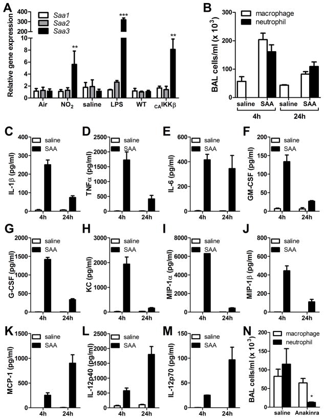
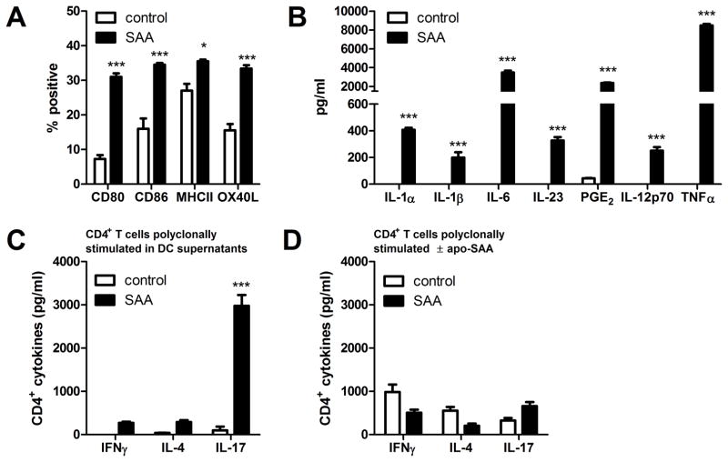
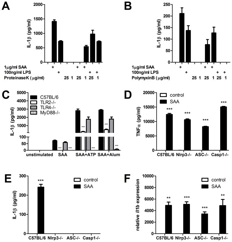
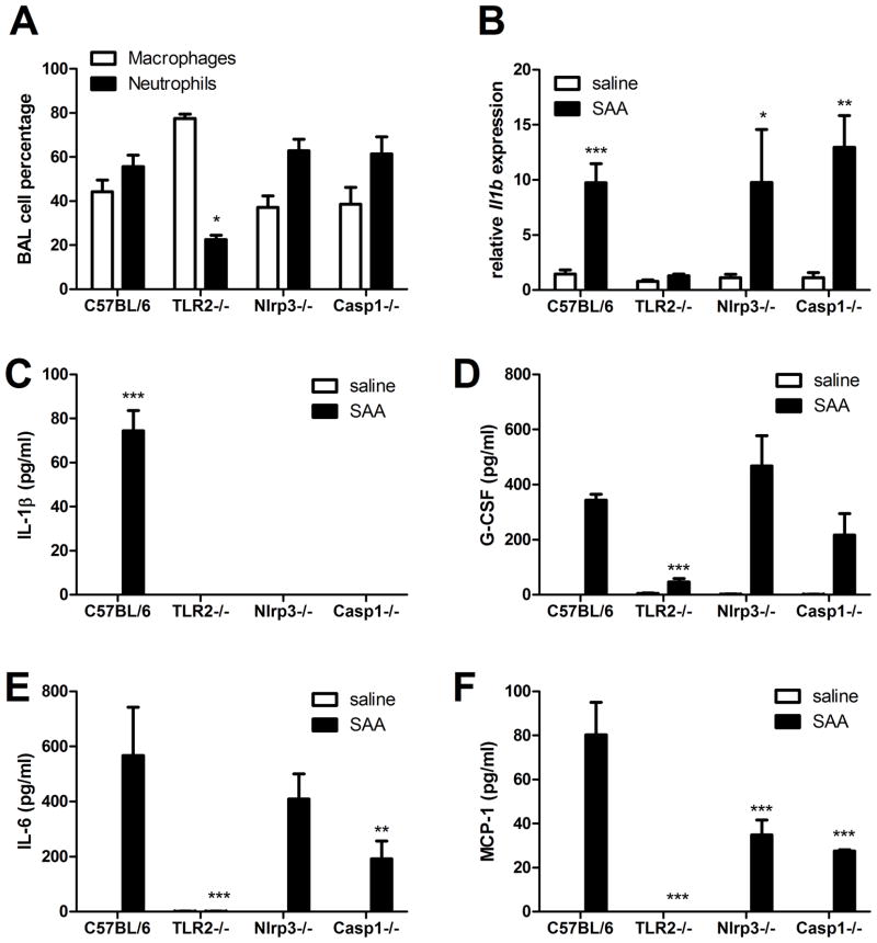
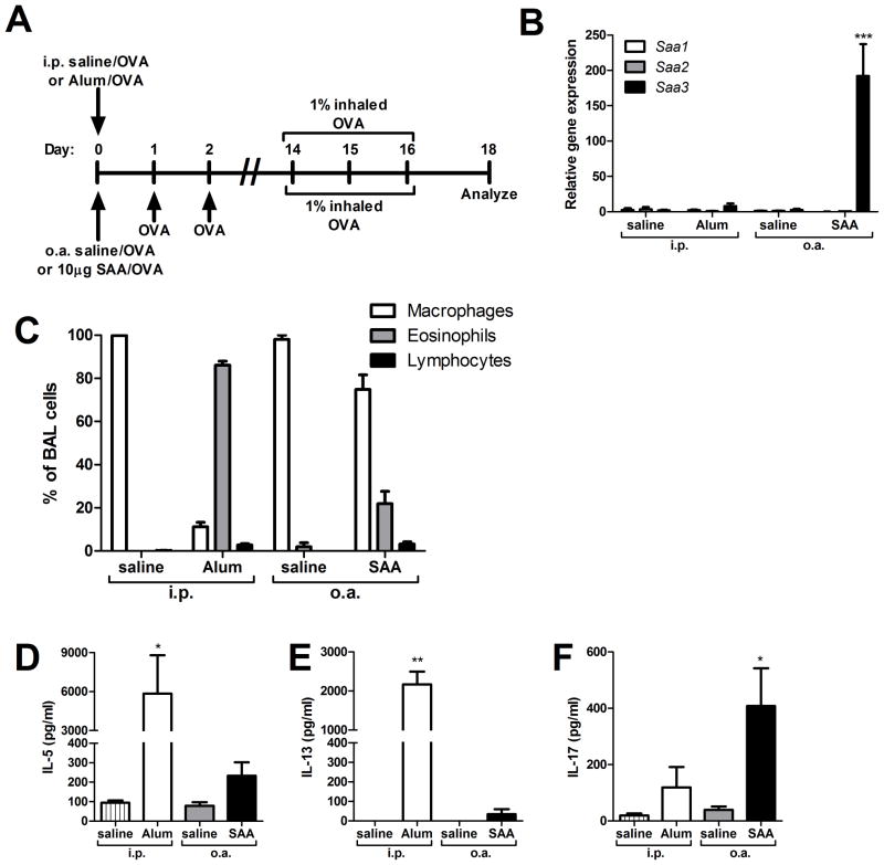
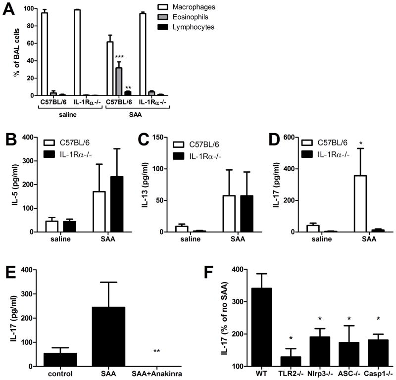
IL-1β is a cytokine critical to several inflammatory diseases in which pathogenic Th17 responses are implicated. Activation of the NLRP3 inflammasome by microbial and environmental stimuli can enable the caspase-1-dependent processing and secretion of IL-1β. The acute-phase protein serum amyloid A (SAA) is highly induced during inflammatory responses, wherein it participates in systemic modulation of innate and adaptive immune responses. Elevated levels of IL-1β, SAA, and IL-17 are present in subjects with severe allergic asthma, yet the mechanistic relationship among these mediators has yet to be identified. In this study, we demonstrate that Saa3 is expressed in the lungs of mice exposed to several mixed Th2/Th17-polarizing allergic sensitization regimens. SAA instillation into the lungs elicits robust TLR2-, MyD88-, and IL-1-dependent pulmonary neutrophilic inflammation. Furthermore, SAA drives production of IL-1α, IL-1β, IL-6, IL-23, and PGE(2), causes dendritic cell (DC) maturation, and requires TLR2, MyD88, and the NLRP3 inflammasome for secretion of IL-1β by DCs and macrophages. CD4(+) T cells polyclonally stimulated in the presence of conditioned media from SAA-exposed DCs produced IL-17, and the capacity of polyclonally stimulated splenocytes to secrete IL-17 is dependent upon IL-1, TLR2, and the NLRP3 inflammasome. Additionally, in a model of allergic airway inflammation, administration of SAA to the lungs functions as an adjuvant to sensitize mice to inhaled OVA, resulting in leukocyte influx after Ag challenge and a predominance of IL-17 production from restimulated splenocytes that is dependent upon IL-1R signaling.


Figures

FIGURE 1
FIGURE 1. SAA is expressed in the lungs during mixed TH2/TH17 allergic sensitization regimens and induces pulmonary inflammation upon inhalational exposure
Quantitative PCR of whole lung for SAA isoforms in mice exposed to NO2, LPS, or in which NF-κB has been activated in the airway epithelium (A). C57BL/6 mice were administered 10 μg of SAA by oropharyngeal aspiration and analyzed 4 and 24 hours later. Bronchoalveolar lavage (BAL) total cell counts were performed by hemocytometer and differential analysis was by cytospin (B). BAL fluid was analyzed by Milliplex assay for IL-1β (C), TNFα (D), IL-6 (E), GM-CSF (F), G-CSF (G), KC (H), MIP-1α (I), MIP-1β (J), MCP-1 (K), IL-12p40 (L), and IL-12p70 (M). Data are representative of three independent experiments. C57BL/6 mice were administered saline or 1 mg Anakinra (n= 3 per group) by subcutaneous injection twice daily beginning one day prior to oropharyngeal aspiration of 10 μg SAA. At 24 hours, BAL total cell counts were performed by hemocytometer and differential analysis was by cytospin (N). * = p<0.05, ** = p<0.005, and *** = p<0.001 compared to control exposures (A) or saline controls (B–N).
FIGURE 2
FIGURE 2. SAA elicits inflammatory mediator production and dendritic cell maturationin vitro
Bone marrow-derived dendritic cells were treated with 1 μg/ml SAA for 16 hours and analyzed for surface markers of maturation (A) and secretion of TH17-polarizing mediators (B). The conditioned media from control or SAA-exposed BMDCs (C) or fresh media with or without SAA (D) was transferred to CD4+ T cells that were polyclonally stimulated with anti-CD3 and anti-CD28. After 96 hours, IFNγ, IL-4, and IL-17A were measured in supernatants. Data are representative of three independent experiments. * = p<0.05, ** = p<0.005, and *** = p<0.001 compared to control exposures.
FIGURE 3
FIGURE 3. SAA-induced IL-1β production requires TLR2, MyD88, and the NLRP3 inflammasome
Cell-free supernatants from bone marrow-derived dendritic cells treated with SAA or LPS in the presence or absence of Proteinase K (A) or Polymyxin B (B) were analyzed by ELISA after 24 hours. Peritoneal exudate macrophages from wild type (C57BL/6), TLR2−/−, TLR4−/−, and MyD88−/− mice were unstimulated or were primed overnight with 1 μg/ml SAA alone or followed by 30 minutes of 5 mM ATP or 8 hours of 500 μg/ml Alum, and supernatants were analyzed for IL-1β secretion (C). Transformed macrophages from wild type (C57BL/6), Nlrp3−/−, ASC−/−, and Caspase-1−/− mice were treated for 24 hours with 1 μg/ml SAA and supernatants were analyzed for TNFα (D) and IL-1β (E) secretion. Il1b expression was measured from wild type (C57BL/6), Nlrp3−/−, ASC−/−, and Caspase-1−/− transformed macrophages that had been untreated or exposed for 4 hours to 1 μg/ml SAA (F). * = p<0.05, ** = p<0.005, and *** = p<0.001 compared to wild type (C) or control exposures (D–F).
FIGURE 4
FIGURE 4. SAA-induced pulmonary inflammation requires TLR2 and involves the NLRP3 inflammasome
Wild type (C57BL/6), TLR2−/−, Nlrp3−/−, and Caspase-1−/− mice were administered 10μg of SAA by oropharyngeal aspiration and analyzed 24 hours later. Bronchoalveolar lavage (BAL) total cell counts were performed by hemocytometer and differential analysis was by cytospin (A). Il1b expression was measured from whole lung by quantitative RT-PCR (B). BAL fluid was analyzed by Milliplex assay for IL-1β (C), G-CSF (D), IL-6 (E), and MCP-1 (F). Data are representative of three independent experiments. * = p<0.05, ** = p<0.005, and *** = p<0.001 compared to wild type (A) or compared to saline (B–F).
FIGURE 5
FIGURE 5. SAA inhalation promotes TH17 allergic sensitization
Mice underwent antigen sensitization via oropharyngeal aspiration (o.a.) with either saline and OVA (saline/OVA) or SAA and OVA (SAA/OVA), or via intraperitoneal (i.p.) injection with either saline and OVA (saline/OVA) or Alum and OVA (Alum/OVA), according to the schema (A). Saa1, Saa2, and Saa3 gene expression in whole lung was measured on day 1, 24 hours after i.p. injection with saline or Alum, or 24 hours after o.a. administration of saline or SAA (B). On day 18, total cell counts from bronchoalveolar lavage (BAL) fluid were performed by hemocytometer and differential analysis was by cytospin (C). On day 18, splenocytes from i.p. saline, i.p. Alum, o.a. saline, and o.a. SAA mice were restimulated in vitro with OVA for 96 hours and IL-5 (D), IL-13 (E), and IL-17A (F) levels in culture media were measured. Data are representative of three independent experiments. * = p<0.05, ** = p<0.005, and *** = p<0.001 compared to saline controls (B, D–F).
FIGURE 6
FIGURE 6. SAA-promoted allergic sensitization and TH17 polarization require IL-1R
C57BL/6 and IL-1Rα−/− mice were antigen-sensitized with saline and OVA (saline) or SAA and OVA (SAA) by o.a., according to the timeline in 5A. On day 18, total and differential cell counts from BAL fluid were performed (A). Splenocytes were restimulated in vitro with OVA for 96 hours and IL-5 (B), IL-13 (C), and IL-17 (D) levels in culture media were measured. Splenocytes from C57BL/6 mice in the presence or absence of SAA and 10 ng/ml Anakinra (E) and splenocytes from C57BL/6, TLR2−/−, NLRP3−/−, ASC−/−, and Casp1−/− (F) mice were polyclonally stimulated for 96 hours with anti-CD3 and anti-CD28 in the presence or absence of 1 μg/ml SAA and IL-17A was measured by ELISA (F). Data are representative of two independent experiments * = p<0.05, ** = p<0.005, and *** = p<0.001 compared to saline controls (A-D), compared to SAA (E) or compared to wild type (F).

References

    1. Glaccum MB, Stocking KL, Charrier K, Smith JL, Willis CR, Maliszewski C, Livingston DJ, Peschon JJ, Morrissey PJ. Phenotypic and functional characterization of mice that lack the type I receptor for IL-1. J Immunol. 1997;159:3364–3371. - PubMed
    1. Masters SL, Dunne A, Subramanian SL, Hull RL, Tannahill GM, Sharp FA, Becker C, Franchi L, Yoshihara E, Chen Z, Mullooly N, Mielke LA, Harris J, Coll RC, Mills KH, Mok KH, Newsholme P, Nunez G, Yodoi J, Kahn SE, Lavelle EC, O’Neill LA. Activation of the NLRP3 inflammasome by islet amyloid polypeptide provides a mechanism for enhanced IL-1beta in type 2 diabetes. Nat Immunol. 11:897–904. - PMC - PubMed
    1. Daheshia M, Yao JQ. The interleukin 1beta pathway in the pathogenesis of osteoarthritis. J Rheumatol. 2008;35:2306–2312. - PubMed
    1. Clutterbuck AL, Mobasheri A, Shakibaei M, Allaway D, Harris P. Interleukin-1beta-induced extracellular matrix degradation and glycosaminoglycan release is inhibited by curcumin in an explant model of cartilage inflammation. Ann N Y Acad Sci. 2009;1171:428–435. - PubMed
    1. Gris D, Ye Z, Iocca HA, Wen H, Craven RR, Gris P, Huang M, Schneider M, Miller SD, Ting JP. NLRP3 plays a critical role in the development of experimental autoimmune encephalomyelitis by mediating Th1 and Th17 responses. J Immunol. 185:974–981. - PMC - PubMed

Publication types

MeSH terms