. 2011 May 27;30(13):2596-609.
doi: 10.1038/emboj.2011.166.

The DNA-binding domain of the Chd1 chromatin-remodelling enzyme contains SANT and SLIDE domains

Affiliations

The DNA-binding domain of the Chd1 chromatin-remodelling enzyme contains SANT and SLIDE domains

Daniel P Ryan et al. EMBO J. .

Abstract

The ATP-dependent chromatin-remodelling enzyme Chd1 is a 168-kDa protein consisting of a double chromodomain, Snf2-related ATPase domain, and a C-terminal DNA-binding domain. Here, we show the DNA-binding domain is required for Saccharomyces cerevisiae Chd1 to bind and remodel nucleosomes. The crystal structure of this domain reveals the presence of structural homology to SANT and SLIDE domains previously identified in ISWI remodelling enzymes. The presence of these domains in ISWI and Chd1 chromatin-remodelling enzymes may provide a means of efficiently harnessing the action of the Snf2-related ATPase domain for the purpose of nucleosome spacing and provide an explanation for partial redundancy between these proteins. Site directed mutagenesis was used to identify residues important for DNA binding and generate a model describing the interaction of this domain with DNA. Through inclusion of Chd1 sequences in homology searches SLIDE domains were identified in CHD6-9 proteins. Point mutations to conserved amino acids within the human CHD7 SLIDE domain have been identified in patients with CHARGE syndrome.


Conflict of interest statement

The authors declare that they have no conflict of interest.

Figures

Figure 1
Figure 1
The C-terminus of Chd1 is required for nucleosome-binding and remodelling activity. (A) Schematic of the domain structure of full-length Chd1 (1–1468) and the C-terminal truncation constructs used in (BD). (B) Nucleosome-binding activity of full-length Chd1 and C-terminal truncations (1–1305ΔC, 1–1010ΔC, and 1–860ΔC). Binding reactions contained 15 nM Cy3-labelled mononucleosomes assembled on 194 bp 601 sequence (Thastrom et al, 1999) DNA (NCP47) and 3.125, 6.25, 12.5, 25, 50, and 100 nM Chd1, 1–1305ΔC, 1–1010ΔC, or 1–860ΔC. Chd1-bound nucleosome species (star) were resolved on native polyacrylamide gels. (C) Nucleosome-sliding activity of recombinant Chd1 and C-terminal truncations. End-positioned mononucleosomes (0.5 pmol) assembled on 201 bp MMTV NucA DNA were incubated with 0.5 nM Chd1, 1–1305ΔC, 1–1010ΔC, or 1–860ΔC for 45 min in the presence of ATP or the non-hydrolysable ATP-analogue ATPγS. Only full-length Chd1 and 1–1305ΔC repositioned nucleosomes to the central location (slower migrating species) in an ATP-dependent manner. (D) DNA- and nucleosome-stimulated ATPase activity is abolished in 1–1010ΔC or 1–860ΔC. ATP hydrolysis catalysed by 5 nM Chd1, 1–1305ΔC, 1–1010ΔC, or 1–860ΔC was monitored in real time using a fluorescent-based phosphate sensor assay in the absence of substrate or in the presence of 100 nM 147 bp DNA, mononucleosomes assembled on 147 bp DNA (NCP), or mononucleosomes assembled on 201 bp DNA (NCP54). Errors bars indicate ±s.d.
Figure 2
Figure 2
Defining the minimal DNA-binding domain of Chd1. (A) Schematic representation of the constructs used to determine the domain boundaries of the C-terminal DNA-binding domain of Chd1. Numbers indicate the boundary residues for each construct. (B) Binding of the constructs described in (A) at 50, 100, and 200 nM to 45 bp Cy3-labelled duplex DNA (50 nM). (C) Binding of Chd1-DBD (55, 110, 220, and 440 nM) to different lengths of Cy3-labelled duplex DNA (50 nM). (D) Binding of Chd1-DBD (55, 110, 220, and 440 nM) to Cy3-labelled nucleosomes (50 nM) assembled on the 601 sequence with (NCP47) or without (NCP) extranucleosomal DNA. Asterisks (*) indicate Chd1-DBD containing complexes.
Figure 3
Figure 3
Structure of S. cerevisiae Chd-DBD. (A) Chd1-DBD sequence aligned to the corresponding regions of Chd1 homologues from S. pombe, C. elegans, D. melanogaster, G. gallus, and H. sapiens. Secondary structure definitions are from the Chd1-DBD crystal structure. The SANT (blue), SLIDE (yellow), helical linker-1 (purple; HL1) and -2 (red; HL2) and β-linker (orange; βL) regions are marked. The sequence alignment is coloured using the ClustalX colouring scheme (Thompson et al, 1997). (B) Cartoon representation of the crystal structure of Chd1-DBD is shown in two orientations. The domains are coloured and marked as denoted in (A).
Figure 4
Figure 4
Comparison of Chd1-DBD with the DmISWI-HAND-SANT-SLIDE (DmISWI-HSS) structure (PDB ID 1OFC). (A) Crystal structures of DmISWI-HSS and Chd1-DBD are shown side by side. The SANT and SLIDE domains in each structure are in the light and dark shade (orange or blue), respectively. (B) Structural superposition of the SLIDE domains of Chd1-DBD and DmISWI-HSS calculated using DALI (RMSD=2.6 Å). The differences in the inserted loop regions between the two structures are marked along with the common inserted helix (α9; also shown in (D)). I, II, and III denote the canonical three helices for SANT/Myb-like domains. (C) Similarly, superpositions of the SANT domains are shown (RMSD=1.6 Å). (D) The shift (∼15 Å) in spacing between the SANT domains of ISWI (orange) and Chd1 (blue) when SLIDE domains are superposed, the other structural elements have been removed for clarity.
Figure 5
Figure 5
The basic surface of Chd1-DBD is necessary for DNA binding. (A) One face of the molecule exhibits a mixture of acidic, basic, and neutral surfaces. The labelled amino acids within the SANT domain comprise an acidic patch on this face. (B) The opposite face of the molecule is almost exclusively positively charged. Key residues involved in DNA binding and underlying structural elements are marked. Electrostatic surface features of Chd1-DBD were calculated using APBS (Baker et al, 2001) (±7 kT/e); blue and red represent positive and negative electrostatic potential, respectively. (C) Wild-type and mutant Chd1-DBD (55, 110, 220, and 440 nM) were tested for binding to Cy3-labelled 20 bp duplex DNA (50 nM). (D) Top-scoring HADDOCK-generated model of Chd1-DBD bound to 20 bp DNA. Upper inset, the interaction between the SANT α1 helix and DNA. Lower inset, the interaction between the SLIDE α10 helix and the minor groove of DNA. Residues in dark blue denote ‘active’ residues in the HADDOCK modelling, residues in cyan are other basic residues found on the binding surface.
Figure 6
Figure 6
The basic surface of Chd1-DBD is important for nucleosome remodelling by Chd1. (A) Full-length wild-type and mutant Chd1 (7.5, 15, 30, and 60 nM) were tested for binding to Cy3-labelled NCP47 nucleosomes (25 nM) (B) Quantification of nucleosome sliding assays using full-length wild-type and mutant Chd1. End-positioned nucleosomes (0.5 pmol) were incubated with Chd1 (0.5 nM) for the times indicated, the products resolved on native polyacrylamide gels and the bands quantitated. Symbols represent the average of at least three experiments and error bars indicate ±s.d. Solid lines are the fits to the data of a single exponential model. (C) ATPase activity of Chd1 mutants (5 nM) in the absence (no substrate) or presence (NCP54) of 100 nM nucleosomes. Data for wild-type Chd1 are taken from Figure 1D for comparison. (D) Yeast complementation assays assessing the ability of full-length wild-type and mutant Chd1 proteins to rescue the temperature sensitive growth defect of an isw1Δ, isw2Δ, chd1Δ triple knockout strain. The triple knockout strain was transformed with single-copy plasmids encoding wild-type or mutant Chd1. Serial dilutions of the transformants and wild-type strain (BMA64) were spotted at equal densities onto selection plates and incubated at 25 or 38°C for 2 days.
Figure 7
Figure 7
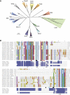
Sequence analysis of Chd1 SANT and SLIDE domains. (A) Unrooted neighbour-joining tree of a multiple sequence alignment of known SANT and Myb domain sequences with SANT domain sequences from Chd1 proteins. Roman numerals refer to the repeat number for proteins containing multiple SANT/Myb domains. *Indicates Myb (II and III) repeats from S. cerevisiae BasI (P22035) and # marks the SANT domain of D. melanogaster dADA2a (Q7KSD8), both outliers for their respective subfamilies. (B) Alignment of SLIDE sequences from Chd1 and ISWI proteins against SLIDE-related sequences from CHD6–9 proteins. Coloured dots indicate amino acids affected by missense (red), non-sense (black), and frameshift (cyan) mutations in the human CHD7 gene of CHARGE syndrome patients. Secondary structure predictions from YASPIN (Lin et al, 2005) and SSPRO (Cheng et al, 2005) structure prediction servers for human CHD9 aligned with the secondary structures of Chd1-DBD and DmISWI SLIDE domains, H=helix, C=coil, E=sheet. Similar predictions were made for the other CHD6–9 sequences. Corresponding helices from the Chd1-DBD structure are indicated.

References

    1. Aasland R, Stewart AF, Gibson T (1996) The SANT domain: a putative DNA-binding domain in the SWI-SNF and ADA complexes, the transcriptional co-repressor N-CoR and TFIIIB. Trends Biochem Sci 21: 87–88 - PubMed
    1. Adams PD, Afonine PV, Bunkoczi G, Chen VB, Davis IW, Echols N, Headd JJ, Hung LW, Kapral GJ, Grosse-Kunstleve RW, McCoy AJ, Moriarty NW, Oeffner R, Read RJ, Richardson DC, Richardson JS, Terwilliger TC, Zwart PH (2010) PHENIX: a comprehensive Python-based system for macromolecular structure solution. Acta Crystallogr D Biol Crystallogr 66: 213–221 - PMC - PubMed
    1. Alen C, Kent NA, Jones HS, O’Sullivan J, Aranda A, Proudfoot NJ (2002) A role for chromatin remodeling in transcriptional termination by RNA polymerase II. Mol Cell 10: 1441–1452 - PubMed
    1. Awad S, Ryan D, Prochasson P, Owen-Hughes T, Hassan AH (2010) The Snf2 homolog Fun30 acts as a homodimeric ATP-dependent chromatin-remodeling enzyme. J Biol Chem 285: 9477–9484 - PMC - PubMed
    1. Bailey S (1994) The Ccp4 suite—programs for protein crystallography. Acta Crystallogr D 50: 760–763 - PubMed

Publication types

MeSH terms

Associated data