. 2011 May 29;474(7353):645-8.
doi: 10.1038/nature10094.

Latent TGF-β binding protein 3 identifies a second heart field in zebrafish

Affiliations

Latent TGF-β binding protein 3 identifies a second heart field in zebrafish

Yong Zhou et al. Nature. .

Abstract

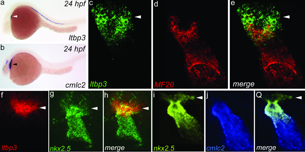
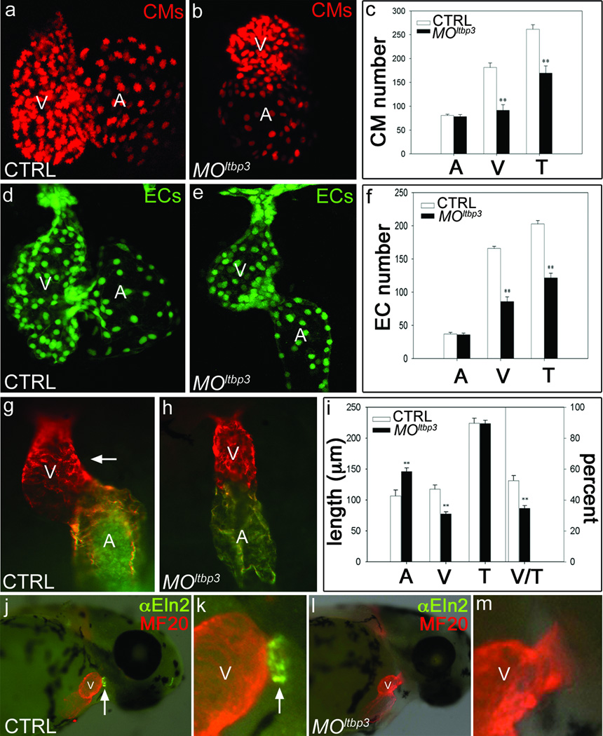
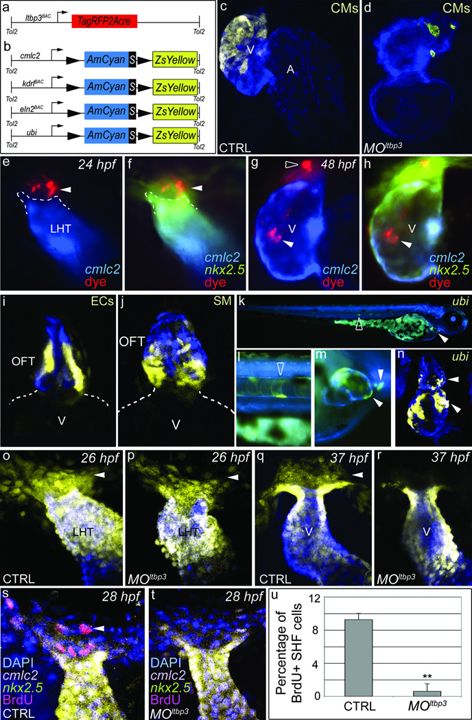
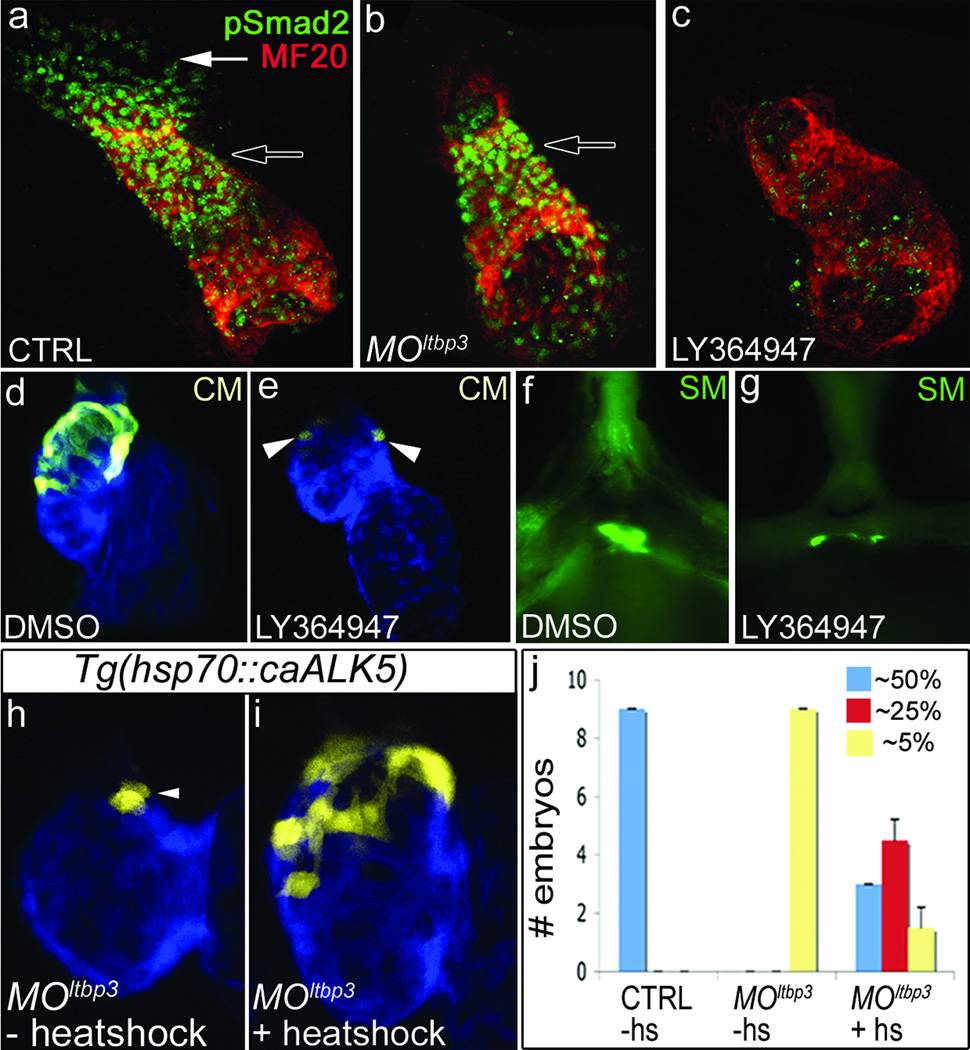
The four-chambered mammalian heart develops from two fields of cardiac progenitor cells distinguished by their spatiotemporal patterns of differentiation and contributions to the definitive heart. The first heart field differentiates earlier in lateral plate mesoderm, generates the linear heart tube and ultimately gives rise to the left ventricle. The second heart field (SHF) differentiates later in pharyngeal mesoderm, elongates the heart tube, and gives rise to the outflow tract and much of the right ventricle. Because hearts in lower vertebrates contain a rudimentary outflow tract but not a right ventricle, the existence and function of SHF-like cells in these species has remained a topic of speculation. Here we provide direct evidence from Cre/Lox-mediated lineage tracing and loss-of-function studies in zebrafish, a lower vertebrate with a single ventricle, that latent TGF-β binding protein 3 (ltbp3) transcripts mark a field of cardiac progenitor cells with defining characteristics of the anterior SHF in mammals. Specifically, ltbp3(+) cells differentiate in pharyngeal mesoderm after formation of the heart tube, elongate the heart tube at the outflow pole, and give rise to three cardiovascular lineages in the outflow tract and myocardium in the distal ventricle. In addition to expressing Ltbp3, a protein that regulates the bioavailability of TGF-β ligands, zebrafish SHF cells co-express nkx2.5, an evolutionarily conserved marker of cardiac progenitor cells in both fields. Embryos devoid of ltbp3 lack the same cardiac structures derived from ltbp3(+) cells due to compromised progenitor proliferation. Furthermore, small-molecule inhibition of TGF-β signalling phenocopies the ltbp3-morphant phenotype whereas expression of a constitutively active TGF-β type I receptor rescues it. Taken together, our findings uncover a requirement for ltbp3-TGF-β signalling during zebrafish SHF development, a process that serves to enlarge the single ventricular chamber in this species.


Figures

Figure 1
Figure 1. ltbp3 and nkx2.5 transcripts mark extra-cardiac cells contiguous to the outflow pole of the zebrafish heart tube
a,b, ltbp3+ cells (white arrowhead) visualized by in situ hybridization at 24 hours post-fertilization (hpf) reside dorsal to the cmlc2+ heart tube (black arrowhead; n=15+ embryos/group). c–e, Heart tube region in 24 hpf embryo co-stained with ltbp3+ riboprobe (green; arrowhead) and a muscle-specific antibody (MF20; red; n=3/3) that recognizes cardiomyocytes. f–h, Heart tube region in 24 hpf embryo co-stained with ltbp3 and nkx2.5 riboprobes highlighting ltbp3+, nkx2.5+ cells (arrowheads) at the outflow pole of the heart tube (n=9/9). i–k, Heart tube region in 26 hpf Tg(nkx2.5::ZsYellow); Tg(cmlc2::CSY) embryo highlighting non-myocardial nkx2.5+ cells (arrowheads).
Figure 2
Figure 2. Knocking down ltbp3 causes multi-lineage cardiovascular defects in the ventricle and OFT
a–f, Cardiomyocyte (CM) and endocardial (EC) cell numbers in control and MOltbp3 atria (A) and ventricles (V) at 72 hpf in cmlc2::dsRed2-nuc and fli1::nEGFP embryos (n=6/group, T=total) g–i, Atrial (A) and ventricular (V) segment lengths in control and MOltbp3 embryos at 36hpf (n=9/group; white arrow highlights rightward looping of the ventricle in control embryos). Error bars in all graphs represent 1 s.d., ** p<0.01. j–m, 60 hpf control and MOltbp3 embryos stained with α-Eln2 antibodies (n=24/24 for WT; 28/31 for MOltbp3) that recognize OFT smooth muscle cell precursor cells (white arrow).
Figure 3
Figure 3. ltbp3+ cells give rise to three cardiovascular lineages in the zebrafish ventricle and OFT
a,b, Schematic of driver (A) and reporter (B) transgenes. c,d, Tg(ltbp3::TagRFP2Acre); Tg(cmlc2::CSY) control and MOltbp3 hearts at 72 hpf (n=>20/group; >90% exhibited ZsYellow distribution shown). e–h, 24 hpf Tg(nkx2.5BAC::ZsYellow); Tg(cmlc2::CSY) embryo injected with tracking dye (arrowheads) in non-myocardial nkx2.5+ region imaged again at 48 hpf. The dye tracked to the distal ventricle (7/16 embryos accurately injected), the myocardial segment of the OFT (5/16; not shown), and both structures (6/16) respectively. Black arrowhead highlights dye that failed to migrate. i, OFT region in Tg(ltbp3::TagRFP2Acre); Tg(kdrl::CSY) embryo at 6 days post fertilization (dpf) [n=16, >50% exhibited ZsYellow fluorescence in OFT endothelial cells (ECs)]. j, OFT region in Tg(ltbp3::TagRFP2Acre); Tg(eln2::CSY) embryo at 7 dpf [n=12, >80% of exhibited ZsYellow fluorescence in OFT smooth muscle (SM) precursors]. k–n, Tg(ltbp3::TagRFP2Acre); Tg(ubi::CSY) embryo at 4 dfp (k–m) and confocal section of the heart on 6 dpf (n) [n=12, >90% of embryos exhibited ZsYellow protein in the distal ventricle (white arrowhead) and OFT (white arrowhead)]. Open arrowheads highlight color switching in the ltbp3+ notochord. o–r, Heart tube regions in control and MOltbp3 Tg(nkx2.5::ZsYellow, cmlc2::CSY) embryos at 26 and 37 hpf. Arrows indicate non-myocardial nkx2.5+ cells present in all experimental groups except MOltbp3 animals at 37hpf (n=18/18 and 12/12 for control and morphants respectively). s-u, 28 hpf control and MOltbp3 Tg(nkx2.5::ZsYellow); Tg(cmlc2::CSY) embryos, processed for DAPI and BrDU staining. The percentages of BrdU+ cells in the ZsYellow+, AmCyan− region are shown in u (n=6 embryos/group, one s.d. is shown, ** p<0.01). V=ventricle, A=atrium, LHT=linear heart tube.
Figure 4
Figure 4. Diminished TGFβ signaling underlies the MOltbp3 cardiovascular phenotypes
a–c, Heart tube regions in 24 hpf control, MOltbp3, and LY364947-treated embryos co-stained with a pSmad2 antibody and a muscle-specific antibody (MF20) that recognizes cardiomyocytes (n =20/20 for a, n=11/11 for b, n=15/15 for c). Black and white arrows highlight the heart tube and extra-cardiac pSmad2+ cells respectively. d, e, 72 hpf hearts in Tg(ltbp3::TagRFP2Acre); Tg(cmlc2::CSY) embryos treated with DMSO or LY364947 (n=8/8 per group); CM=cardiomyocytes. f, g, OFT region in 60 hpf embryos treated with DMSO or LY364947 and immunostained for Eln2 epitopes (n=20/20). SM=smooth muscle precursors. h–j, Control and MOltbp3 Tg(ltbp3::TagRFP2Acre); Tg(cmlc2::CSY); Tg(hsp70::caALK5) embryos were heat shocked (+hs) or not (−hs) and evaluated visually at 72hpf. Graph in (k) indicates the number of embryos in each experimental group that expressed ZsYellow protein in the distal ~50% (full rescue), ~25% (partial rescue), or ~5% (no rescue) of the ventricle. Shown in (j) is a heart that is partially rescued.

References

    1. Dyer LA, Kirby ML. The role of secondary heart field in cardiac development. Dev Biol. 2009;336(2):137–144. - PMC - PubMed
    1. Rochais F, Mesbah K, Kelly RG. Signaling pathways controlling second heart field development. Circ Res. 2009;104(8):933–942. - PubMed
    1. Vincent SD, Buckingham ME. How to make a heart: the origin and regulation of cardiac progenitor cells. Curr Top Dev Biol. 2010;90:1–41. - PubMed
    1. Olson EN. Gene regulatory networks in the evolution and development of the heart. Science. 2006;313(5795):1922–1927. - PMC - PubMed
    1. Cai CL, et al. Isl1 identifies a cardiac progenitor population that proliferates prior to differentiation and contributes a majority of cells to the heart. Dev Cell. 2003;5(6):877–889. - PMC - PubMed

Publication types

Associated data