. 2011 Aug;118(3):365-78.
doi: 10.1111/j.1471-4159.2011.07330.x. Epub 2011 Jun 17.

Pituitary adenylate cyclase-activating peptide induces long-lasting neuroprotection through the induction of activity-dependent signaling via the cyclic AMP response element-binding protein-regulated transcription co-activator 1

Affiliations
Free PMC article

Pituitary adenylate cyclase-activating peptide induces long-lasting neuroprotection through the induction of activity-dependent signaling via the cyclic AMP response element-binding protein-regulated transcription co-activator 1

Paul S Baxter et al. J Neurochem. 2011 Aug.
Free PMC article

Abstract

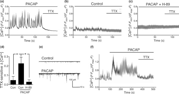
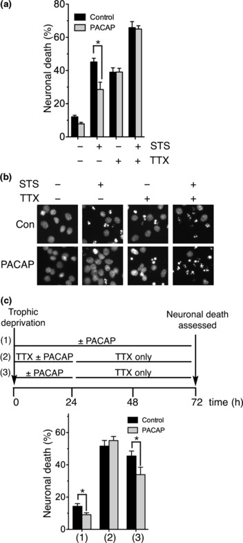
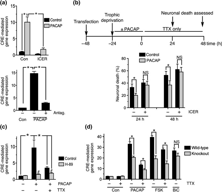
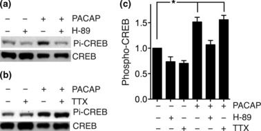
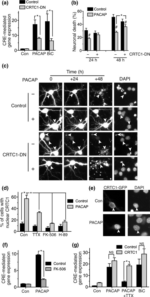
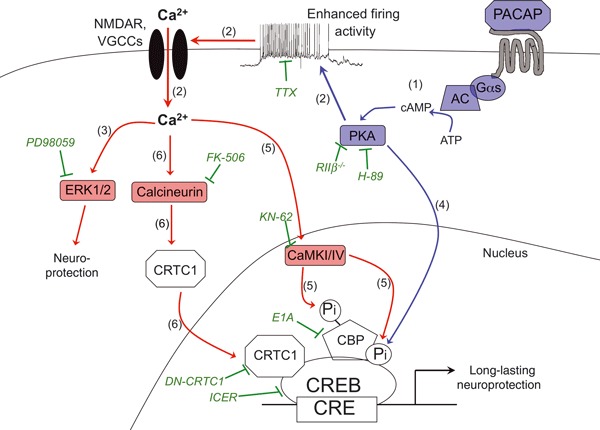
Pituitary adenylate cyclase-activating peptide (PACAP) is a neuroprotective peptide which exerts its effects mainly through the cAMP-protein kinase A (PKA) pathway. Here, we show that in cortical neurons, PACAP-induced PKA signaling exerts a major part of its neuroprotective effects indirectly, by triggering action potential (AP) firing. Treatment of cortical neurons with PACAP induces a rapid and sustained PKA-dependent increase in AP firing and associated intracellular Ca(2+) transients, which are essential for the anti-apoptotic actions of PACAP. Transient exposure to PACAP induces long-lasting neuroprotection in the face of apoptotic insults which is reliant on AP firing and the activation of cAMP response element (CRE) binding protein (CREB)-mediated gene expression. Although direct, activity-independent PKA signaling is sufficient to trigger phosphorylation on CREB's activating serine-133 site, this is insufficient for activation of CREB-mediated gene expression. Full activation is dependent on CREB-regulated transcription co-activator 1 (CRTC1), whose PACAP-induced nuclear import is dependent on firing activity-dependent calcineurin signaling. Over-expression of CRTC1 is sufficient to rescue PACAP-induced CRE-mediated gene expression in the face of activity-blockade, while dominant negative CRTC1 interferes with PACAP-induced, CREB-mediated neuroprotection. Thus, the enhancement of AP firing may play a significant role in the neuroprotective actions of PACAP and other adenylate cyclase-coupled ligands.


Figures

Fig. 1
Fig. 1
PACAP enhances AP firing in cortical neurons. (a–d) Pre-treatment with PACAP (10 nM PACAP-27 here and throughout the study) causes an increase in AP firing-dependent Ca2+ transients. Neurons were treated where indicated with PACAP ± H-89 (10 μM). After 2 h, the neurons were subjected to Fluo-3 Ca2+ imaging studies (see Methods for details) to monitor the size of Ca2+ transients in the different stimulation conditions. TTX (1 μM) was added where indicated to determine the extent to which the observed Ca2+ transients were because of action potential firing. Example traces are shown: black line indicates the mean Ca2+ concentration within a field of cells, and the grey shaded region indicates ± SEM of the Ca2+ concentration within that field. Free Ca2+ concentrations were calculated from fluorescence signal (F) according to the equation [Ca2+] = Kd(FFmin)/(FmaxF), and expressed as a multiple of the Kd of Fluo-3 (which is approximately 315 nM). (d) shows quantification of data shown in (a–c), that is, quantification of the difference in mean amplitude of [Ca2+] before and after 1 μM TTX treatment. In order to quantitate the effect of PACAP on firing activity-induced [Ca2+] influx, the mean [Ca2+] 30 s before and 30 s after TTX treatment was calculated in either control neurons or neurons treated with PACAP ± H-89. For each cell, the degree of TTX-sensitive Ca2+ changes was calculated as the difference between mean [Ca2+] before and after TTX treatment. For each condition, 60 cells were analysed within six independent experiments (*p < 0.05). (e) Example trace of a whole-cell voltage-clamp recording of a control and PACAP-treated cortical neurons. PACAP causes an increase in burst-like activity, consistent with the Ca2+ imaging data. (f) Ca2+ imaging of acute PACAP treatment, a typical example trace is shown representative of six independent experiments.
Fig. 2
Fig. 2
PACAP promotes resistance to apoptotic stimuli which is dependent on AP firing. (a,b) PACAP protects against staurosporine-induced cell death, but not in the presence of TTX. Neurons were treated with PACAP in the presence or absence of TTX 24 h and 1 h before treatment with 100 nM staurosporine. After a further 24 h, the cells were then fixed and DAPI stained and death was measured by counting pyknotic and non-pyknotic nuclei (*p < 0.05, n = 4); (b) shows example pictures. (c) PACAP-induced AP firing protects against trophic deprivation and promotes long lasting neuroprotection. At t = 0, the neurons were placed in trophically-deprived medium and given one of the three treatment regimes outlined in the upper schematic (1–3). At t = 72 h, cells were fixed, DAPI stained and levels of neuronal death analysed (*p < 0.05, n = 3).
Fig. 3
Fig. 3
PACAP induces CRE-dependent gene expression, which is neuroprotective, and relies on AP firing. (a) Upper-PACAP induces CRE-mediated gene expression. Neurons were transfected with a CRE-Firefly luciferase vector, pTK renilla transfection control and vectors encoding either ICER1 or control (β-globin). See Methods section for exact quantities used. At 24 h post-transfection, neurons were treated with PACAP and luciferase expression was measured after a further 4 h. CRE-Firefly luciferase activity was normalised to Renilla control (*p < 0.05, n = 3). Lower-Effect of the PACAP antagonist (Antag, PACAP6–38, 1 μM) on PACAP induction of CRE-luciferase (*p < 0.05, n = 3). (b) PACAP mediated long-lasting neuroprotection depends on activation of CRE-mediated gene expression. Upper panel illustrates the experimental protocol. Briefly, neurons expressing GFP plus either ICER1 or β-globin control were treated ± PACAP 24 h post-transfection and then all cells were placed in TTX-containing medium after a further 24 h, at which point images of GFP-expressing neurons were taken (t = 0 in the upper schematic). The fate of these cells was then monitored at 24 and 48 h after this medium change. 250–400 cells were analysed per treatment across six cultures within three independent experiments. (*p < 0.05). (c) PACAP induced CRE-dependent gene expression is dependent on AP firing. Neurons were treated with PACAP where indicated for 4 h; all other drugs were added 1 h beforehand (*p < 0.05, #p < 0.05 comparing H-89 with control for that particular PACAP/TTX condition, n = 7). (d) PACAP and forskolin-induced activation of CRE-mediated gene expression is disrupted in RIIβ-deficient neurons: both AP firing-dependent and independent components. Forskolin was used at 5 μM. For comparison is an illustration of the RIIβ-independence of CRE activation triggered by promoting AP firing by network disinhibition through treatment with the GABAA receptor blocker bicuculline (50 μM) plus 250 μM 4-aminopyridine, which is a PKA-independent way of inducing AP firing (Papadia et al. 2005) (*p < 0.05, n = 6).
Fig. 4
Fig. 4
PACAP-mediated induction of serine-133 CREB phosphorylation does not require AP firing. (a–c) PACAP induces phosphorylation of CREB at serine-133 in a TTX-insensitive, PKA-dependent manner. Neurons were pre-treated with TTX or H-89 and then treated for 15 min with PACAP. Protein was harvested and subject to western analysis for phospho-CREB (see Methods, normalized in all cases to total CREB, *p < 0.05, n = 4, example blots are shown). (a) and (b) show example westerns and (c) shows quantitation of phospho-CREB levels (normalized to total CREB).
Fig. 5
Fig. 5
PACAP induces nuclear translocation of CRTC1, necessary for the AP firing-dependent component of CREB activation. (a) CRTC1 dominant negative inhibits PACAP mediated activation of CREB. Neurons were transfected with CRE-luciferase, pTK-Renilla and vectors encoding either a CRTC1 dominant negative mutant or control (β-globin). Neurons were stimulated PACAP or bicuculline plus 4-AP (BiC) (*p < 0.05, n = 4). (b,c) PACAP mediated long-lasting neuroprotection depends on CRTC1. The experimental protocol is the same as that illustrated schematically in Fig. 3(b). Briefly, neurons expressing GFP plus either CRTC1-DN (dominant negative) or β-globin control were treated ± PACAP 24 h post-transfection and then all cells were placed in TTX-containing medium after a further 24 h, at which point images of GFP-expressing neurons were taken. The fate of these cells was then monitored at 24 and 48 h after this medium change (*p < 0.05, paired T-test, n = 3; #p < 0.05, paired T-test comparing control to PACAP within each condition/timepoint). (c) shows example pictures. Scale bar = 20 μm. (d,e) PACAP induces CRTC1 nuclear translocation via activity-dependent calcineurin signaling. Neurons were transfected with a vector encoding GFP-tagged CRTC1. At 24 h post-transfection, neurons were treated with 20 ng/mL leptomycin B for 30 min to block nuclear export [to enable import to be observed more clearly (Kovacs et al. 2007)], plus the indicated inhibitors (1 μM TTX, 10 μM H-89 or 10 μM FK-506) and then PACAP added for 30 min prior to fixing of the cells and analysing localisation of GFP-CRTC1 in 400–800 cells per treatment (*p < 0.05, n = 4–8). (e) shows example pictures. (f) PACAP-induced activation of CRE-mediated gene expression requires the Ca2+-dependent phosphatase calcineurin. Where used, FK-506 was added 1 h prior to PACAP stimulation (*p < 0.05, n = 4). (g) CRTC1 over-expression rescues the inhibition of PACAP-mediated CRE activation by TTX. Neurons were transfected with CRE-luciferase, pTK-Renilla and either vectors encoding CRTC1 or β-globin control. 24 h post-transfection the neurons were stimulated with PACAP ± TTX or bicuculline + 4-AP (BiC) as indicated. Over-expression of CRTC1 does not further enhance CRE activation by BiC or PACAP, suggesting that levels are not limiting, however, it strongly enhances levels induced by PACAP in the presence of TTX (*p < 0.05, n = 4).
Fig. 6
Fig. 6
Schematic illustration of the role of activity-dependent Ca2+ signaling in PACAP-mediated neuroprotection. Activation of PACAP receptors leads to activation of PKA via the classical G-protein-adenylate cyclase (AC)-cAMP: pathway (1). PKA activation causes an increase in synaptic strength and/or neuronal excitability leading to a strong increase in levels of action potential firing which in turn triggers intracellular Ca2+ influx, likely through synaptic receptors (e.g. NMDA receptors) or voltage-gated Ca2+ channels (VGCCs): pathway (2). Activation of long-lasting neuroprotection by PACAP requires induction of gene expression mediated by the transcription factor CREB. CREB phosphorylation on serine-133 can be triggered directly by PKA in an AP firing-independent manner: pathway (3). However, this is insufficient to fully activate CREB-mediated gene expression. A key Ca2+/activity-dependent pathway involves CRTC1 nuclear translocation through activation of the Ca2+-dependent phosphatase calcineurin: pathway (4). Blue arrows and molecules indicate AP firing activity-independent events, while red arrows and molecules highlight the events dependent on AP firing. The pharmacological and genetic inhibitors of the various pathways used in this study are shown in green.

References

    1. Al-Mubarak B, Soriano FX, Hardingham GE. Synaptic NMDAR activity suppresses FOXO1 expression via a cis-acting FOXO binding site: FOXO1 is a FOXO target gene. Channels (Austin, Tex) 2009;3:233–238. - PMC - PubMed
    1. Atlasz T, Szabadfi K, Kiss P, et al. Pituitary adenylate cyclase activating polypeptide in the retina: focus on the retinoprotective effects. Ann. N Y Acad. Sci. 2010;1200:128–139. - PubMed
    1. Aubert N, Falluel-Morel A, Vaudry D, et al. PACAP and C2-ceramide generate different AP-1 complexes through a MAP-kinase-dependent pathway: involvement of c-Fos in PACAP-induced Bcl-2 expression. J. Neurochem. 2006;99:1237–1250. - PubMed
    1. Bading H, Greenberg ME. Stimulation of protein tyrosine phosphorylation by NMDA receptor activation. Science. 1991;253:912–914. - PubMed
    1. Banke TG, Bowie D, Lee H, Huganir RL, Schousboe A, Traynelis SF. Control of GluR1 AMPA receptor function by cAMP-dependent protein kinase. J. Neurosci. 2000;20:89–102. - PMC - PubMed

Publication types

MeSH terms