. 2011 Aug;78(2):476.e1-8.
doi: 10.1016/j.urology.2011.03.024. Epub 2011 May 31.

Functional, metabolic, and morphologic characteristics of a novel rat model of type 2 diabetes-associated erectile dysfunction

Affiliations

Functional, metabolic, and morphologic characteristics of a novel rat model of type 2 diabetes-associated erectile dysfunction

Maarten Albersen et al. Urology. 2011 Aug.

Abstract

Objectives: To conduct a pilot study to investigate functional, metabolic, and penile morphologic changes in a novel model of lean DM2. Erectile dysfunction (ED) is a frequent sequela in patients with type 2 diabetes mellitus (DM2).

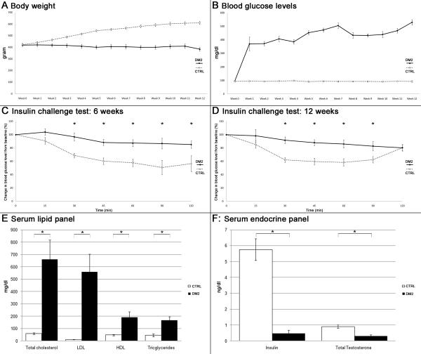
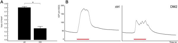
Methods: Eight rats received a high-fat diet and 2 weeks later, 2 intraperitoneal injections of streptozotocin (STZ, 30 mg/kg). Five age-matched rats served as controls. Insulin challenge tests were performed at 6 and 12 weeks after induction of DM2. At 12 weeks, erectile function was tested by measurement of intracavernous pressure (ICP) increase upon cavernous nerve stimulation. Penile tissue and serum samples were harvested for histology and biochemistry, respectively.

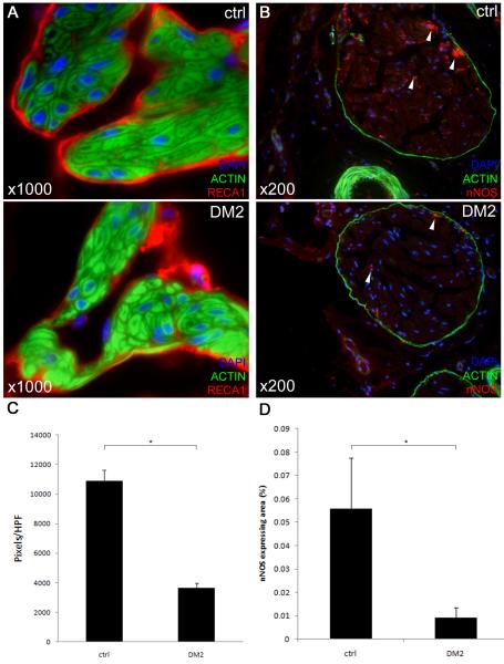
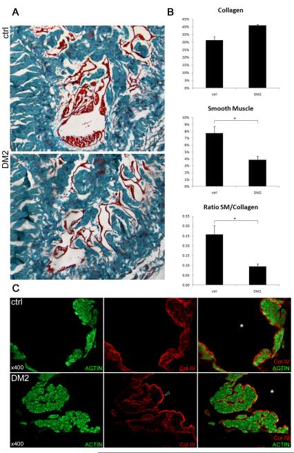
Results: A lean DM2 model was established as demonstrated by decreased insulin resistance, elevated nonfasting plasma glucose levels, hyperlipidemia, and decreased insulin concentration in the absence of obesity. ICP/mean arterial pressure was significantly decreased in DM2 animals (0.29) compared with controls (0.81). Expression of neuronal nitric oxide synthase and rat endothelial cell antigen-1, and the smooth muscle/collagen ratio were significantly decreased in the penis of DM2 animals.

Conclusions: We propose an inexpensive nongenetic animal model of lean DM2-associated ED. Microanatomical changes in the erectile tissue that reflect an advanced stage of the disease were observed.


Figures

Fig. 1
Fig. 1. Growth curves, glucose metabolism and serum biochemistry
A: Graph depicts growth curves of the experimental animals. Body weight was measured weekly starting at the day of the first STZ injection. B: Postprandial blood glucose levels were measured weekly starting at the day of the first STZ injection. C,D: Insulin challenge test carried out 6 and 12 weeks following the first STZ injection. *: p < 0.05. E: Levels of lipids on rat serum biochemistry evaluation. *: p < 0.05. F: Levels of insulin and total testosterone in rat serum determined by ELISA. *: p < 0.05.
Fig. 2
Fig. 2. Erectile function upon cavernous nerve electrostimulation
A: The effects of induction of DM2 on ratio of ICP/mean arterial pressure (MAP). *: p < 0.05. B: Representative ICP recordings per group. Note the spiky appearance of the plateau phase of the erection in diabetic rats, indicative of inability to maintain erection. The red bar represents an electrical stimulation of the cavernous nerve with a duration of 50 s.
Fig. 3
Fig. 3. Changes in the endothelium and dorsal penile nerves in the diabetic rat penis
A: Representative images of endothelial lining of sinusoids. Rat penis was stained with Alexa-488-conjugated phalloidin and with Alexa-594-conjugated anti-RECA1 antibody. Original magnification ×1000. Note the ‘patchy’ appearance of the endothelial lining in diabetic penile sinusoids whereas the endothelial lining is continuous in normal rats. B: Representative images of rat penile dorsal nerves. Rat penis was stained with Alexa-488-conjugated phalloidin and with Alexa-594-conjugated anti-nNOS antibody. Original magnification ×200. Note the significant decrease of nNOS positive nerve fibers, which are located mainly in the periphery of the dorsal nerve (arrows). C: Quantification of RECA1-staining in pixels/HPF, *: p < 0.001. D: Quantification of nNOS-staining in dorsal penile nerves, *: p < 0.001.
Fig 4
Fig 4. Smooth muscle and extracellular matrix in the corpus cavernosum
A: Representative pictures of the rat corpus cavernosum stained with Masson’s trichrome. Red color represents mainly smooth muscle, blue stains collagen fibers. Original magnification ×40. B: Histomorphometric analysis of the content of the corpus cavernosum. Upper and middle panel: collagen content and smooth muscle content in percentage of total intratunical area, lower panel: ratio smooth muscle (%) / collagen (%).*: p < 0.05. C: Rat penis was stained with Alexa-488-conjugated phalloidin and with Alexa-594-conjugated anti-collagen-IV antibody. Original magnification ×400. Representative high-power images of smooth muscle bundles lining a cavernosal sinusoid (*). Upper panel: normal rat, lower panel: diabetic rat. Note the somewhat denser expression of the sinusoidal subendothelial basement membrane in the diabetic rat sinus (arrowhead).

References

    1. Giugliano F, Maiorino M, Bellastella G, et al. Determinants of erectile dysfunction in type 2 diabetes. Int J Impot Res. 2010;22(3):204–9. - PubMed
    1. Chitaley K. Type 1 and Type 2 diabetic-erectile dysfunction: same diagnosis (ICD-9), different disease? J Sex Med. 2009;6(Suppl 3):262–8. - PubMed
    1. Hidalgo-Tamola J, Chitaley K. Review type 2 diabetes mellitus and erectile dysfunction. J Sex Med. 2009;6(4):916–26. - PubMed
    1. Costa C, Soares R, Castela A, et al. Increased endothelial apoptotic cell density in human diabetic erectile tissue--comparison with clinical data. J Sex Med. 2009;6(3):826–35. - PubMed
    1. Wingard C, Fulton D, Husain S. Altered penile vascular reactivity and erection in the Zucker obese-diabetic rat. J Sex Med. 2007;4(2):348–62. discussion 362-3. - PubMed

Publication types